File:National Museum of Health and Medicine.jpg
National_Museum_of_Health_and_Medicine.jpg (800 × 600 pixels, file size: 201 KB, MIME type: image/jpeg)
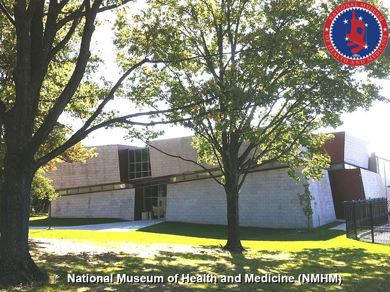
National Museum of Health and Medicine (USA)
The National Museum of Health and Medicine (NMHM) new building opened on September 15, 2011 located on Linden Lane in Silver Spring, Montgomery County, Maryland (USA).
The museum contains the Human Developmental Anatomy Centre Collections that include the historic Carnegie Collection of human embryos.
Human Developmental Anatomy Center Collections
The following information are excerpts from the Guide to Collections (2014) p198 - 201. This section of the guide was previously revised by Elizabeth Lockett and Emily Wilson (2009 and 2012).
Arey-Dapeña Pediatric Pathology Collection
- 24 boxes, 5 binders.
- Finding aid, restricted, digitized.
- Over 6,000 lantern slides of various pediatric pathologies, represented by both gross and histological images, created by Marie (Molly) Valdes-Dapeña at the University of Miami.
- All slides have been digitized and a searchable database is available.
AFIP Sudden Infant Death Collection
- A collection of wet tissue blocks sealed in plastic, glass slides, and detailed case histories documenting cases of Sudden Infant Death Syndrome.
- Materials were originally acquired in the 1970s and 1980s at the National Institute of Child Health and Human Development.
Birth Defects Encyclopedia
- Records and photographs used to compile the Birth Defects Encyclopedia by Mary Louise Buyse.
- Mary Louise Buyse, M.D., who served as the president and editor-in-chief of the Center for Birth Defects Information Services
- A printed copy of the 1892-page encyclopedia and an electronic file used to produce the print version are included in the collection.
Buyse M. (1980). Center for Birth Defects Information Services. Birth Defects Orig. Artic. Ser. , 16, 83-91. PMID: 7448380
Carnegie Collection Of Embryology
- This collection consists principally of serial sections of normal human embryo development in the first eight weeks, as started by the Carnegie Institution of Washington’s Department of Embryology in 1914.
- The collection now forms the core of the Human Developmental Anatomy Center.
- Extensive collateral materials include case histories, photographs, micrographs, models in various media, and comparative materials (mostly rhesus).
- Reprints from the Carnegie Contributions to Embryology, other reprints relating to embryology, films, and personal documents are also available.
- This collection incorporates other embryo collections, such as the Davis Orthopedic Development and Elizabeth Mapelsden Ramsey Collections.
- Partially digitized.
Davis Orthopedic Development Collection
- Part of the Carnegie Collection of Embryology added while curated at the University of California, Davis.
- A large collection of glass slides charting bone growth and development in the human.
- Slides are grouped by structure starting with the head and moving down through the torso and out the extremities.
Hertig Videos
- Six VHS tapes of lectures given by Dr. Arthur T. Hertig, an embryologist who worked extensively with the Carnegie materials.
- The tapes include two each of: trophoblastic disease, malignant disease of the uterus, and ovarian tumors.
- See also OHA 189, Hertig Collection.
Cornell School Of Veterinary Med. Embryological Collection
* About 150 boxes, 12 binders.
- From the Cornell School of Veterinary Medicine in Ithaca, NY this collection includes human, rat, mouse and guinea pig embryos.
- There is still a large collection of embryonic material at Cornell.
Gaensler Pulmonary Pathology Collection
- A collection of radiographic images and case histories of lung diseases, primarily asbestosis.
- Transferred to the Archives in November 2008 as OHA 163.55.
Hooker-Humphrey Collection
- Originally compiled at the University of Pittsburgh by Davenport Hooker (1887-1965) and Tryphena Humphrey (1902-1971), this collection came to the Museum via the University of Alabama.
- This collection of human and comparative material is stained to highlight nervous system development.
- Sizes of specimens range from 3mm to 310mm. For those wishing to study the development of the human nervous system from birth through maturity to old age.
- This collection is a bridge between the Carnegie Collection focusing on the first eight weeks of development and the neonatal to adult material in the Yakovlev-Haleem Collection.
- http://brainmuseum.org
Charles Sedgwick Minot Embryological Collection
- Embryos from the Harvard School of Medicine, as well as drawings and photographs of the embryos.
- A large collection of reprints, printed lectures, class syllabi, and theses on embryology and related topics.
- The reprint collection was started by Charles S. Minot (1852-1914) in the 1800s and added to through the 1960s.
- The reprint collection also includes personal papers and research notes from Charles Wislocki.
- Finding aid, arranged?
Elizabeth Mapelsden Ramsey Collection
- Reprints, personal papers, correspondence, research notes, and artifacts from Elizabeth Mapelsden Ramsey.
- Ramsey was a former curator of the Carnegie Collection, as well as a researcher, lecturer, and teacher.
- The collection is subsumed by the Carnegie Collection of Embryology.
Sensenig Collection
- A small set of comparative material on glass slides from the University of Alabama, compiled by E. Carl Sensenig.
- Arrived along with the Hooker Humphrey comparative materials.
Patten- Burdi Collection
- This is a collection of sectioned embryos and fetuses, focused on the second and third trimesters.
- It includes histology sections, reprints, paste-ups for various editions of Patten's Embryology, 2 whole mounts in celloidin, records, and photographic materials.
- Alphonse Burdi was the last curator of the collection at University of Michigan and organized its donation to the museum.
- The Embryology Research Collection at Michigan was established by embryologists George Streeter and G. Carl Huber in the early 1900s with a mission to collect and describe the morphogenesis of human embryos at critical stages in prenatal life.
- For about twenty years up to 1957, the Collection grew in numbers under the leadership of Professor Bradley M. Patten whose primary interests were in heart and cardiovascular development.
- Human Embryology, by Bradley M. Patten, Ph.D. New York: McGraw-Hill Book Company, 1st edn. 1946; 2nd edn. 1953; 3rd edn. 1968
Berkowitz Cleft Palate
- The collection of orthodontist Samuel Berkowitz includes panoramic x-rays, cephalometrics, patient records, dental casts, digital records, and educational materials of his longitudinal studies on treatment of cleft lip and palate.
- Dr. Berkowitz was a Clinical Professor of Pediatrics and Surgery associated with the South Florida Craniofacial Anomalies Program at the University of Miami School of Medicine and is now active in developing teaching materials on cleft palate.
Selected Reference Forsberg A. (1974). [Frequency of broken endodontic files]. Tandlakartidningen , 66, 8-9. PMID: 4524205
- Search Pubmed: Berkowitz S (Author)
Koering Collection
- A former student of George Corner, Dr. Marilyn J. Koering (1938-2008) studied follicle and ovary development.
- The collection consists of histology slides (mostly rhesus), photographs, micrographs, personal reprints, reprints supporting research, and research notes.
Stanford Mouse
- A teaching set collection of mouse embryo development.
Ob-Gyn Lantern Slides
- A collection of lantern slides used in teaching obstetrics and gynecology dating from 1915-1940.
- Much of the contained data pertaining to New York clinics (e.g., Berwind and Bellevue).
- The set represents popular contemporary topics in the field.
George Washington University Medical School Fetal Development Collection
- A collection of whole embryos and fetuses, with associated records, used for teaching and display by GWU Medical College, created and donated by Frank D. Allan.
Richardson Pediatric Slide Collection
- Notebooks of 35mm slides of lecture text and clinical pictures of various pediatric pathologies and diseases from the pediatric practice of Martyn E. Richardson.
- Topics include musculoskeletal syndromes; neurosensory; blood and RE; cardiovascular; endocrine; genitor-urinary; infectious diseases; drug company slides; newborn; gastrointestinal; nutrition; skin; respiratory; allergy; collagen diseases; growth and development; and child abuse.
- 10 binders and 71 loose pages. Arranged, unrestricted.
Lockard Reprint Collection
- Reprint collection of embryologist Isabel Lockard. May also contain photographs.
- 6 boxes. No finding aid, unarranged, inactive, unrestricted.
- Isabel Lockard, Ph.D., (1915 -2011) - neuroanatomist at Georgetown University (1947-1952). At Medical College of South Carolina’s Department of Anatomy as Assistant Professor (1952). Second full-time female faculty member in the College of Medicine, and the first neuroanatomist at the Medical University to retain tenure in the position over the long term. She was a Professor 1969-1985). Retirement and appointed as Professor Emeritus in 1985.
- Links: Waring Historical Library
Kimmel Collection
- Histology slides of 25 human embryo specimens from research by Donald L. Kimmel and Elizabeth Moyer, Temple University, 1940s-50s.
- Search PubMed: KIMMEL DL (Author)
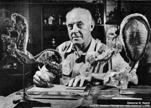
Osborne O. Heard Collection
- This collection is primarily a record of anatomical modeler Osborne O. Heard's (1890-1983) work while at the Carnegie Institute of Washington's Department of Embryology.
- It includes correspondence, early photographs of the department, and Mr. Heard.
- Artwork includes sketches by James Didusch and mechanical drawings of various devices designed by Mr. Heard for the CIW and other departments at John Hopkins. Dates 1919-1989.
Johns Hopkins Medical School Lantern Slides
- A collection of 2 boxes consisting of 500+ lantern slides used at Johns Hopkins University Medical School for teaching embryology, 1920-2009.
- Includes a number of images from the Carnegie Collection of Embryology.
- Arrived along with models that were originally from, and are now re-incorporated into, the Carnegie Collection.
Reference
Original image http://commons.wikimedia.org/wiki/File:NMHM_20111006c.jpg cropped and relabeled.
Copyright
This file is licensed under the Creative Commons Attribution 3.0 Unported license.
Cite this page: Hill, M.A. (2024, April 27) Embryology National Museum of Health and Medicine.jpg. Retrieved from https://embryology.med.unsw.edu.au/embryology/index.php/File:National_Museum_of_Health_and_Medicine.jpg
- © Dr Mark Hill 2024, UNSW Embryology ISBN: 978 0 7334 2609 4 - UNSW CRICOS Provider Code No. 00098G
File history
Click on a date/time to view the file as it appeared at that time.
| Date/Time | Thumbnail | Dimensions | User | Comment | |
|---|---|---|---|---|---|
| current | 11:32, 16 April 2015 |  | 800 × 600 (201 KB) | Z8600021 (talk | contribs) | ==National Museum of Health and Medicine (USA)== |
You cannot overwrite this file.
File usage
The following page uses this file:


