File:Nanagas1925-fig01e.jpg
Original file (600 × 765 pixels, file size: 52 KB, MIME type: image/jpeg)
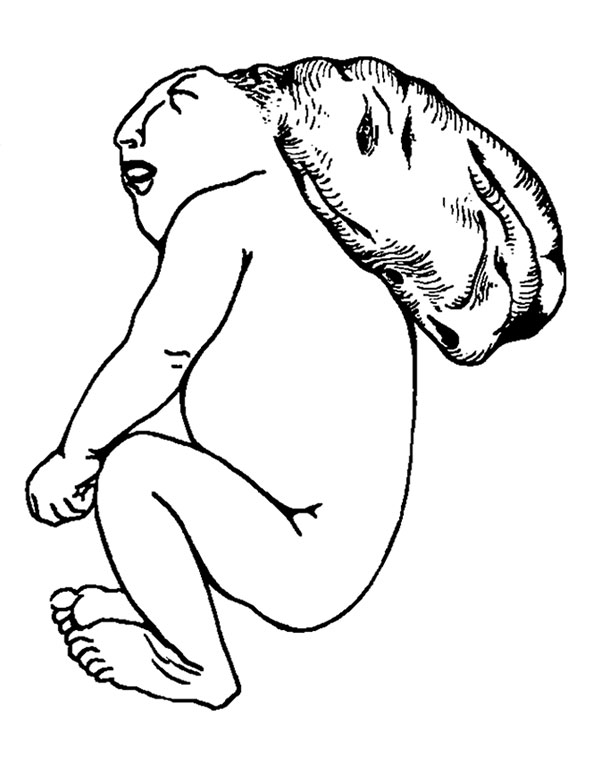
Fig. 1d. Exocephalic Acranius
As the term signifies, specimens of this type show hemiae cerebri, usually pendulous and attached to the back of the head. The cranial vault is likewise absent as in all the preceding groups of cases. The covering of the flattened head is formed by an ordinary scalp continuous with the skin supporting the herniated encephalon. Only one example of this type of exencephalus was encountered in our series. The pendulous mass, suspended by a stalk, was cystic in character, and the hemispheres of the brain were filled with fluid.
The appearance of the face in all these various types of anencephalus is very striking. The eyes are markedly protruded, with a prominent bulging of the upper eyelids — a condition obviously due to the receded superciliary ridges produced by the disappearance of the forehead. The nose appears enlarged. The tongue in many of these cases is considerably protruded.
The neck is abnormally short; so much so, that the head appears to be attached directly to the shoulders and upper end of the thorax. In fetuses with open vertebral canals the head is hyperextended with the face looking upward and its base is set firmly against the back of the shoulders. The trunk region in these specimens is relatively much shorter than in other types. This is partly due to the marked anterior curvature of the vertebral column.
| Historic Disclaimer - information about historic embryology pages |
|---|
 |
- Links: Fig 1. Anencephalus types | Fig 1a. anencephalic acranius | Fig 1b. anencephalic craniorhachischisis | Fig 1c. microcephalic acrauius | Fig 1d. microcephalic craniorhachischisis | Fig 1e. exocephalic acranius | Fig 16. anencephalic and normal fetuses | Historic Embryology Papers | Neural Abnormalities | Folic Acid and Neural Tube Defects | Skull Development
Reference
Nañagas JC. A comparison of the growth of the body dimensions of anencephalic human fetuses with normal fetal growth as determined by graphic analysis and empirical formulae. (1925) American J. Anatomy. 455-494.
Cite this page: Hill, M.A. (2024, April 27) Embryology Nanagas1925-fig01e.jpg. Retrieved from https://embryology.med.unsw.edu.au/embryology/index.php/File:Nanagas1925-fig01e.jpg
- © Dr Mark Hill 2024, UNSW Embryology ISBN: 978 0 7334 2609 4 - UNSW CRICOS Provider Code No. 00098G
File history
Click on a date/time to view the file as it appeared at that time.
| Date/Time | Thumbnail | Dimensions | User | Comment | |
|---|---|---|---|---|---|
| current | 12:23, 16 September 2015 | | 600 × 765 (52 KB) | Z8600021 (talk | contribs) | {{Nanagas1925 figures}} |
You cannot overwrite this file.