File:Muscle fiber types.jpg: Difference between revisions
m (→Type II fibres) |
mNo edit summary |
||
| (5 intermediate revisions by the same user not shown) | |||
| Line 1: | Line 1: | ||
==Muscle | ==Muscle Fibre Types== | ||
Muscle | Muscle fibre types identified by ATPase staining. Myosin binds and hydrolyzes ATP during force generation. | ||
===Type I fibres | ===Type I fibres=== | ||
Red muscles contain predominantly (but not exclusively) red muscle cells. Red muscle fibres are comparatively thin and contain large amounts of myoglobin and mitochondria. Red fibres contain an isoform of myosin with low ATPase activity, i.e. the speed with which myosin is able to use up ATP. Contraction is therefore slow. Red muscles are used when sustained production of force is necessary, e.g. in the control of posture. | Red muscles contain predominantly (but not exclusively) red muscle cells. Red muscle fibres are comparatively thin and contain large amounts of myoglobin and mitochondria. Red fibres contain an isoform of myosin with low ATPase activity, i.e. the speed with which myosin is able to use up ATP. Contraction is therefore '''slow'''. Red muscles are used when sustained production of force is necessary, e.g. in the control of posture. | ||
===Type II fibres=== | ===Type II fibres=== | ||
White muscle cells, which are predominantly found in white muscles, are thicker and contain less myoglobin. ATPase activity of the myosin isoform in white fibres is high, and contraction is fast. Type IIA fibres (red) contain many mitochondria and are available for both sustained activity and short-lasting, intense contractions. Type IIB/IIX fibres (white) contain only few mitochondria. They are recruited in the case of rapid accelerations and short lasting maximal contraction. Type IIB/IIX fibres rely on anaerobic glycolysis to generate the ATP needed for contraction | White muscle cells, which are predominantly found in white muscles, are thicker and contain less myoglobin. ATPase activity of the myosin isoform in white fibres is high, and contraction is '''fast'''. | ||
* '''Type IIA''' fibres (red) contain many mitochondria and are available for both sustained activity and short-lasting, intense contractions. | |||
* '''Type IIB/IIX''' fibres (white) contain only few mitochondria. They are recruited in the case of rapid accelerations and short lasting maximal contraction. Type IIB/IIX fibres rely on anaerobic glycolysis to generate the ATP needed for contraction. | |||
{{Muscle fibre type table}} | |||
{{Muscle fibre type collapsible table}} | |||
:'''Links:''' [[Musculoskeletal_System_-_Muscle_Development|Muscle Development]] | |||
{{Blue Histology}} | {{Blue Histology}} | ||
[[Category:Histology]] [[Category:Muscle]] | [[Category:Histology]] [[Category:Muscle]] | ||
Latest revision as of 12:47, 7 October 2015
Muscle Fibre Types
Muscle fibre types identified by ATPase staining. Myosin binds and hydrolyzes ATP during force generation.
Type I fibres
Red muscles contain predominantly (but not exclusively) red muscle cells. Red muscle fibres are comparatively thin and contain large amounts of myoglobin and mitochondria. Red fibres contain an isoform of myosin with low ATPase activity, i.e. the speed with which myosin is able to use up ATP. Contraction is therefore slow. Red muscles are used when sustained production of force is necessary, e.g. in the control of posture.
Type II fibres
White muscle cells, which are predominantly found in white muscles, are thicker and contain less myoglobin. ATPase activity of the myosin isoform in white fibres is high, and contraction is fast.
- Type IIA fibres (red) contain many mitochondria and are available for both sustained activity and short-lasting, intense contractions.
- Type IIB/IIX fibres (white) contain only few mitochondria. They are recruited in the case of rapid accelerations and short lasting maximal contraction. Type IIB/IIX fibres rely on anaerobic glycolysis to generate the ATP needed for contraction.
| Fibre Type | Type I fibres | Type II a fibres | Type II x fibres | Type II b fibres |
|---|---|---|---|---|
| Contraction time | Slow | Moderately Fast | Fast | Very fast |
| Size of motor neuron | Small | Medium | Large | Very large |
| Resistance to fatigue | High | Fairly high | Intermediate | Low |
| Activity Used for | Aerobic | Long-term anaerobic | Short-term anaerobic | Short-term anaerobic |
| Maximum duration of use | Hours | <30 minutes | <5 minutes | <1 minute |
| Power produced | Low | Medium | High | Very high |
| Mitochondrial density | High | High | Medium | Low |
| Capillary density | High | Intermediate | Low | Low |
| Oxidative capacity | High | High | Intermediate | Low |
| Glycolytic capacity | Low | High | High | High |
| Major storage fuel | Triglycerides | Creatine phosphate, glycogen | Creatine phosphate, glycogen | Creatine phosphate, glycogen |
| Myosin heavy chain, human genes |
MYH7 | MYH2 | MYH1 | MYH4 |
| Muscle Fibre Type | ||||
|---|---|---|---|---|
| Fibre Type | Type I fibres | Type II a fibres | Type II x fibres | Type II b fibres |
| Contraction time | Slow | Moderately Fast | Fast | Very fast |
| Size of motor neuron | Small | Medium | Large | Very large |
| Resistance to fatigue | High | Fairly high | Intermediate | Low |
| Activity Used for | Aerobic | Long-term anaerobic | Short-term anaerobic | Short-term anaerobic |
| Maximum duration of use | Hours | <30 minutes | <5 minutes | <1 minute |
| Power produced | Low | Medium | High | Very high |
| Mitochondrial density | High | High | Medium | Low |
| Capillary density | High | Intermediate | Low | Low |
| Oxidative capacity | High | High | Intermediate | Low |
| Glycolytic capacity | Low | High | High | High |
| Major storage fuel | Triglycerides | Creatine phosphate, glycogen | Creatine phosphate, glycogen | Creatine phosphate, glycogen |
| Myosin heavy chain, human genes |
MYH7 | MYH2 | MYH1 | MYH4 |
| Links: Muscle Development | Muscle Development Timeline | ||||
- Links: Muscle Development
Links: Histology | Histology Stains | Blue Histology images copyright Lutz Slomianka 1998-2009. The literary and artistic works on the original Blue Histology website may be reproduced, adapted, published and distributed for non-commercial purposes. See also the page Histology Stains.
Cite this page: Hill, M.A. (2024, April 26) Embryology Muscle fiber types.jpg. Retrieved from https://embryology.med.unsw.edu.au/embryology/index.php/File:Muscle_fiber_types.jpg
- © Dr Mark Hill 2024, UNSW Embryology ISBN: 978 0 7334 2609 4 - UNSW CRICOS Provider Code No. 00098G
File history
Click on a date/time to view the file as it appeared at that time.
| Date/Time | Thumbnail | Dimensions | User | Comment | |
|---|---|---|---|---|---|
| current | 15:47, 15 September 2009 | 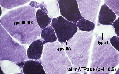 | 400 × 250 (49 KB) | S8600021 (talk | contribs) | Image source: UWA Blue Histology http://www.lab.anhb.uwa.edu.au/mb140/CorePages/Muscle/Muscle.htm#SKELETAL Copyright Lutz Slomianka 1998-2009. The literary and artistic works (excluding the UWA logo) on this web-site may be reproduced, adapted, publish |
You cannot overwrite this file.
File usage
The following 8 pages use this file: