File:Mouse pineal E15 to E21 neuroepithelium 01.jpg

Original file (1,280 × 2,123 pixels, file size: 1.1 MB, MIME type: image/jpeg)
Mouse Pineal Gland (E15-21)
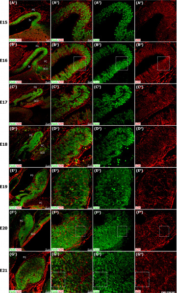
The pineal gland develops from neuroepithelial cells that express the transcription factor Pax6 and the intermediate filament vimentin.
Panels display confocal microscopy of immunolabeled sagittal sections of rat pineal gland (PG) from embryonic day (E) 15 to E21. Both male and female rat embryos were used.
Panels A1-G1 show expression of Pax6 (green) and vimentin (VIM, red) in pineal precursor cells at low magnification.
Panels A2-G2 display the same structures at higher magnification. Panels A3-G3 and A4-G4 show Pax6 and vimentin expression for each stage of development, respectively. Pineal organogenesis begins around E15 as an evagination of the neuroepithelium in the dorsal diencephalon that is densely populated by Pax6-expressing cells (green). The developing PG becomes a tubular extension at E16. The orientation of Pax6/VIM+ cells is radial at these stages. At E17 the pineal neuroepithelium begins to fold and fuses at the midline. After fusion of the neuroepithelium, double immunolabeled rosette-like structures are visible in the E18-E21 developing PG. At E21 the PG has developed into a recognizable globular structure. Inset boxes are shown at higher magnification in Fig 3. (A1-G1) 20x; scale bar: 75 μm. (A2-G4) 60x; scale bar: 25 μm. PC, posterior commissure. SCO, subcommissural organ. 3v, third ventricle.
- Links: Pineal Development | Glial Development | Mouse Development
Reference
Ibañez Rodriguez MP, Noctor SC & Muñoz EM. (2016). Cellular Basis of Pineal Gland Development: Emerging Role of Microglia as Phenotype Regulator. PLoS ONE , 11, e0167063. PMID: 27861587 DOI.
Ibañez Rodriguez MP, Noctor SC, Muñoz EM (2016) Cellular Basis of Pineal Gland Development: Emerging Role of Microglia as Phenotype Regulator. PLoS ONE 11(11): e0167063. https://doi.org/10.1371/journal.pone.0167063
Editor: Serge Nataf, Universite Claude Bernard Lyon 1, FRANCE
Received: July 28, 2016; Accepted: November 8, 2016; Published: November 18, 2016
Copyright
© 2016 Ibañez Rodriguez et al. This is an open access article distributed under the terms of the Creative Commons Attribution License, which permits unrestricted use, distribution, and reproduction in any medium, provided the original author and source are credited.
Fig 1. https://doi.org/10.1371/journal.pone.0167063.g001 adjusted in size and PMID label added.
Cite this page: Hill, M.A. (2024, April 27) Embryology Mouse pineal E15 to E21 neuroepithelium 01.jpg. Retrieved from https://embryology.med.unsw.edu.au/embryology/index.php/File:Mouse_pineal_E15_to_E21_neuroepithelium_01.jpg
- © Dr Mark Hill 2024, UNSW Embryology ISBN: 978 0 7334 2609 4 - UNSW CRICOS Provider Code No. 00098G]
File history
Click on a date/time to view the file as it appeared at that time.
| Date/Time | Thumbnail | Dimensions | User | Comment | |
|---|---|---|---|---|---|
| current | 12:20, 22 May 2017 |  | 1,280 × 2,123 (1.1 MB) | Z8600021 (talk | contribs) | The pineal gland develops from neuroepithelial cells that express the transcription factor Pax6 and the intermediate filament vimentin. Panels display confocal microscopy of immunolabeled sagittal sections of rat pineal gland (PG) from embryonic day (... |
You cannot overwrite this file.
File usage
The following page uses this file: