File:Mouse Purkinje neuron-01.jpg
Mouse_Purkinje_neuron-01.jpg (498 × 578 pixels, file size: 152 KB, MIME type: image/jpeg)
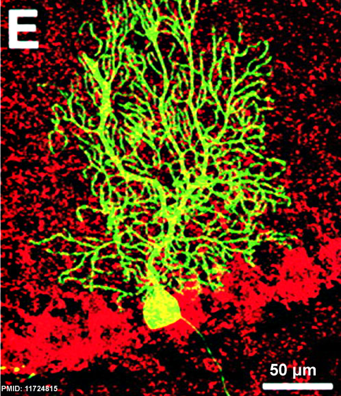
Mouse Purkinje neuron
This GFP labelled mouse Purkinje cell shows the extensive dendritic tree (top), soma or cell body (centre) and the thin axon (bottom).
Figure legend text - Confocal microscopic analysis of BM-derived Purkinje cells. 12 mo after transplantation of BM cells from transgenic mice that ubiquitously express GFP, donor-derived Purkinje cells were visualized by confocal laser–scanning microscopy. The GFP fluorescence of an engrafted neuron.
GFP-marked neuron is visible in the layer of GAD-expressing Purkinje cells (E, overlay of the maximum intensity projections of GFP and Texas red fluorescence visualizing GAD immunoreactivity).
Scale bar 50 μm
Reference
<pubmed>11724815</pubmed>| PMC2150878 | J Cell Biol.
Copyright
Rockefeller University Press - Copyright Policy This article is distributed under the terms of an Attribution–Noncommercial–Share Alike–No Mirror Sites license for the first six months after the publication date (see http://www.jcb.org/misc/terms.shtml). After six months it is available under a Creative Commons License (Attribution–Noncommercial–Share Alike 4.0 Unported license, as described at https://creativecommons.org/licenses/by-nc-sa/4.0/ ). (More? Help:Copyright Tutorial)
Figure 5. panel E cropped and resized.
File history
Click on a date/time to view the file as it appeared at that time.
| Date/Time | Thumbnail | Dimensions | User | Comment | |
|---|---|---|---|---|---|
| current | 13:25, 17 October 2012 | | 498 × 578 (152 KB) | Z8600021 (talk | contribs) | ==Mouse Purkinje neuron== Confocal microscopic analysis of BM-derived Purkinje cells. 12 mo after transplantation of BM cells from transgenic mice that ubiquitously express GFP, donor-derived Purkinje cells were visualized by confocal laser–scanning mi |
You cannot overwrite this file.
File usage
The following 3 pages use this file: