File:Mouse E18.5 cochlea sem04.jpg
Original file (905 × 535 pixels, file size: 85 KB, MIME type: image/jpeg)
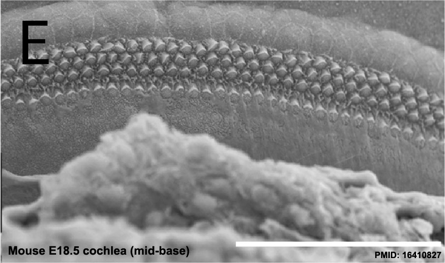
Mouse Embryo (E18.5) Cochlea SEM
Note the inner hair cells (single row) and outer hair cells (3 rows).
The stereocilia can be seen arranged in a shallow "V" shape similar to an "organ pipe" arrangement.
- Links: Cochlea overview SEM | Base region SEM | Mid-base and Apex region SEM | Mid-base region SEM | Mid-base hair cells SEM | Inner Ear Development | Hearing | Mouse Development
Reference
Kiernan AE, Xu J & Gridley T. (2006). The Notch ligand JAG1 is required for sensory progenitor development in the mammalian inner ear. PLoS Genet. , 2, e4. PMID: 16410827 DOI.
Copyright
© 2006 Kiernan et al. This is an open-access article distributed under the terms of the Creative Commons Attribution License, which permits unrestricted use, distribution, and reproduction in any medium, provided the original author and source are credited.
Figure 5. doi:10.1371/journal.pgen.0020004.g005 (images cropped and relabelled from full figure)
Cite this page: Hill, M.A. (2024, April 27) Embryology Mouse E18.5 cochlea sem04.jpg. Retrieved from https://embryology.med.unsw.edu.au/embryology/index.php/File:Mouse_E18.5_cochlea_sem04.jpg
- © Dr Mark Hill 2024, UNSW Embryology ISBN: 978 0 7334 2609 4 - UNSW CRICOS Provider Code No. 00098G
File history
Click on a date/time to view the file as it appeared at that time.
| Date/Time | Thumbnail | Dimensions | User | Comment | |
|---|---|---|---|---|---|
| current | 14:30, 17 February 2013 | | 905 × 535 (85 KB) | Z8600021 (talk | contribs) | Mouse E18.5 cochlea sem03.jpg {{Mouse cochlea sem links}} |
You cannot overwrite this file.
File usage
There are no pages that use this file.