File:Mouse-hatching blastocyst.jpg: Difference between revisions
From Embryology
| Line 7: | Line 7: | ||
Original image name: Figure 3. Viability of Embryos Post-assay. (image extracted from full figure) | Original image name: Figure 3. Viability of Embryos Post-assay. (image extracted from full figure) | ||
==Reference== | ===Reference=== | ||
<pubmed>20405021</pubmed>| [http://www.plosone.org/article/info%3Adoi%2F10.1371%2Fjournal.pone.0010160 PLoS One.] | <pubmed>20405021</pubmed>| [http://www.plosone.org/article/info%3Adoi%2F10.1371%2Fjournal.pone.0010160 PLoS One.] | ||
Latest revision as of 11:42, 18 June 2011
Mouse Hatching Blastocyst
Hatched blastocyst stage ( post-fertilization Day 4.5).
Scale bar 50 µm.
Original image name: Figure 3. Viability of Embryos Post-assay. (image extracted from full figure)
Reference
<pubmed>20405021</pubmed>| PLoS One.
© 2010 Valley et al. This is an open-access article distributed under the terms of the Creative Commons Attribution License, which permits unrestricted use, distribution, and reproduction in any medium, provided the original author and source are credited.
File history
Click on a date/time to view the file as it appeared at that time.
| Date/Time | Thumbnail | Dimensions | User | Comment | |
|---|---|---|---|---|---|
| current | 16:12, 7 October 2010 | 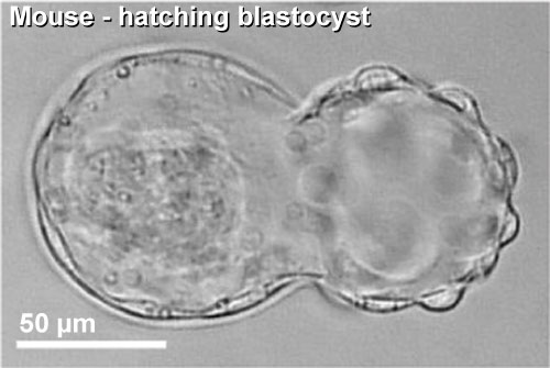 | 500 × 335 (27 KB) | S8600021 (talk | contribs) | ==Mouse Hatching Blastocyst== Hatched blastocyst stage ( post-fertilization Day 4.5). Scale bar 50 µm. Original image name: Figure 3. Viability of Embryos Post-assay. extracted from full figure) ==Reference== <pubmed>20405021</pubmed>| [http://www.p |
You cannot overwrite this file.
File usage
The following 3 pages use this file: