File:Mouse- zona pellucida 01.jpg: Difference between revisions
No edit summary |
|||
| Line 1: | Line 1: | ||
==Light and Electron Micrographs of the Mouse Zona Pellucida== | ==Light and Electron Micrographs of the Mouse Zona Pellucida== | ||
A - light micrograph (Nomarski-differential interference contrast) of sperm bound to the egg zona pellucida. | '''A''' - light micrograph (Nomarski-differential interference contrast) of sperm bound to the egg zona pellucida. | ||
Scale bar ∼ 13 μm. | Scale bar ∼ 13 μm. | ||
B - scanning electron micrograph of the egg zona pellucida. | '''B''' - scanning electron micrograph of the egg zona pellucida. | ||
:(taken from Familiari, G., Relucenti, M., Heyn, R., Micara, G., and Correr, S. (2006) Microscopy Res. Tech. 69, 415–426 with permission for JBC paper). | :(taken from Familiari, G., Relucenti, M., Heyn, R., Micara, G., and Correr, S. (2006) Microscopy Res. Tech. 69, 415–426 with permission for JBC paper). | ||
Scale bar ∼ 200 nm. | Scale bar ∼ 200 nm. | ||
==Reference== | ===Reference=== | ||
<pubmed>18539589</pubmed>| [http://www.jbc.org/content/283/36/24285.long J Biol Chem.] | <pubmed>18539589</pubmed>| [http://www.jbc.org/content/283/36/24285.long J Biol Chem.] | ||
| Line 16: | Line 16: | ||
{{ | {{JBC}} | ||
Original file name: FIGURE 1. http://www.jbc.org/content/283/36/24285/F1.large.jpg | |||
[[Category:Mouse]] [[Category:Oocyte]] [[Category:Spermatozoa]] [[Category:Zona Pellucida]] | [[Category:Mouse]] [[Category:Oocyte]] [[Category:Spermatozoa]] [[Category:Zona Pellucida]] | ||
Revision as of 23:23, 17 January 2013
Light and Electron Micrographs of the Mouse Zona Pellucida
A - light micrograph (Nomarski-differential interference contrast) of sperm bound to the egg zona pellucida.
Scale bar ∼ 13 μm.
B - scanning electron micrograph of the egg zona pellucida.
- (taken from Familiari, G., Relucenti, M., Heyn, R., Micara, G., and Correr, S. (2006) Microscopy Res. Tech. 69, 415–426 with permission for JBC paper).
Scale bar ∼ 200 nm.
Reference
<pubmed>18539589</pubmed>| J Biol Chem.
© the American Society for Biochemistry and Molecular Biology. "Other parties are welcome to copy, distribute, transmit and adapt the work — at no cost and without permission — for noncommercial use as long as they attribute the work to the original source using the citation above." JBC Copyright Permission Policy
Original file name: FIGURE 1. http://www.jbc.org/content/283/36/24285/F1.large.jpg
File history
Click on a date/time to view the file as it appeared at that time.
| Date/Time | Thumbnail | Dimensions | User | Comment | |
|---|---|---|---|---|---|
| current | 08:55, 25 February 2011 | 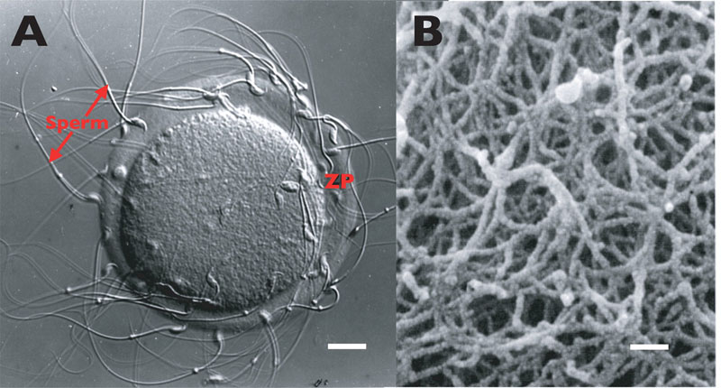 | 800 × 430 (83 KB) | S8600021 (talk | contribs) | ==Light and Electron Micrographs of the Mouse Zona Pellucida== A - light micrograph (Nomarski-differential interference contrast) of sperm bound to the egg ZP. Scale bar ∼ 13 μm. B - scanning electron micrograph of the egg ZP :(taken from Familia |
You cannot overwrite this file.
File usage
The following 2 pages use this file: