File:Mouse- hippocampus dentate granule cells.jpg

Original file (1,000 × 671 pixels, file size: 130 KB, MIME type: image/jpeg)
Mouse Hippocampus
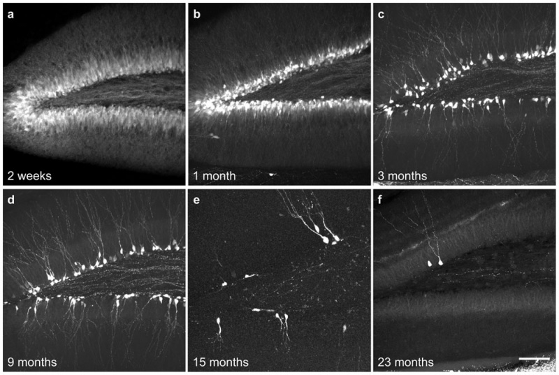
The developmental changes of GFP+ newborn dentate granule cells.
Z stack confocal images show the developmental changes of GFP+ newborn neuron numbers at 2 weeks, 1 month, 3 months, 9 months, 15 months and 23 months.
Scale bar is 100 µm.
- "Neurogenesis in the adult hippocampus is an important form of structural plasticity in the brain. Here we report a line of BAC transgenic mice (GAD67-GFP mice) that selectively and transitorily express GFP in newborn dentate granule cells of the adult hippocampus. These GFP+ cells show a high degree of colocalization with BrdU-labeled nuclei one week after BrdU injection and express the newborn neuron marker doublecortin and PSA-NCAM. Compared to mature dentate granule cells, these newborn neurons show immature morphological features: dendritic beading, fewer dendritic branches and spines. These GFP+ newborn neurons also show immature electrophysiological properties: higher input resistance, more depolarized resting membrane potentials, small and non-typical action potentials. The bright labeling of newborn neurons with GFP makes it possible to visualize the details of dendrites, which reach the outer edge of the molecular layer, and their axon (mossy fiber) terminals, which project to the CA3 region where they form synaptic boutons. GFP expression covers the whole developmental stage of newborn neurons, beginning within the first week of cell division and disappearing as newborn neurons mature, about 4 weeks postmitotic. Thus, the GAD67-GFP transgenic mice provide a useful genetic tool for studying the development and regulation of newborn dentate granule cells."
- Links: hippocampus
Reference
Zhao S, Zhou Y, Gross J, Miao P, Qiu L, Wang D, Chen Q & Feng G. (2010). Fluorescent labeling of newborn dentate granule cells in GAD67-GFP transgenic mice: a genetic tool for the study of adult neurogenesis. PLoS ONE , 5, . PMID: 20824075 DOI.
Copyright
© 2010 Zhao et al. This is an open-access article distributed under the terms of the Creative Commons Attribution License, which permits unrestricted use, distribution, and reproduction in any medium, provided the original author and source are credited.
Original file name: Figure 5. Journal.pone.0012506.g005.jpg
Cite this page: Hill, M.A. (2024, April 27) Embryology Mouse- hippocampus dentate granule cells.jpg. Retrieved from https://embryology.med.unsw.edu.au/embryology/index.php/File:Mouse-_hippocampus_dentate_granule_cells.jpg
- © Dr Mark Hill 2024, UNSW Embryology ISBN: 978 0 7334 2609 4 - UNSW CRICOS Provider Code No. 00098G
File history
Click on a date/time to view the file as it appeared at that time.
| Date/Time | Thumbnail | Dimensions | User | Comment | |
|---|---|---|---|---|---|
| current | 07:44, 7 November 2010 |  | 1,000 × 671 (130 KB) | S8600021 (talk | contribs) | ==Mouse Hippocampus== The developmental changes of GFP+ newborn dentate granule cells. Z stack confocal images show the developmental changes of GFP+ newborn neuron numbers at 2 weeks, 1 month, 3 months, 9 months, 15 months and 23 months. Scale bar is |
You cannot overwrite this file.
File usage
The following page uses this file: