File:Mesenchymal cell cytoplasmic extensions 01.jpg
Original file (1,000 × 846 pixels, file size: 241 KB, MIME type: image/jpeg)
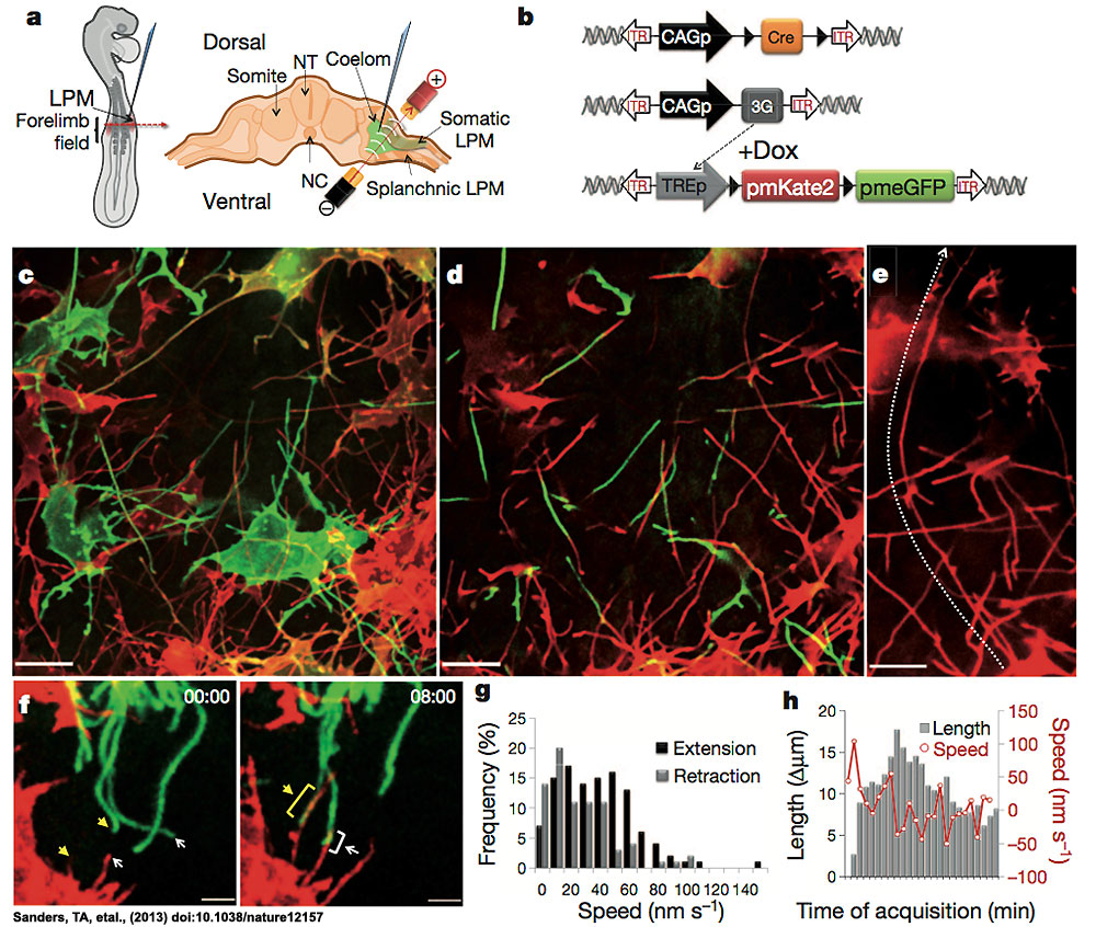
Mesenchymal cells of the developing limb bud possess long and highly dynamic cytoplasmic extensions
a - Left, HH14 chick embryo indicating the site of DNA injection. The red dashed line indicates the cross-sectional plane. Right, microinjected DNA in the coelom is shown in green; the electrodes position is indicated (LPM). NC, notochord; NT, neural tube.
b - Diagram of piggyBac transposon system resulting in integration of transposon inverted terminal repeat (ITR)-flanked expression cassettes by piggyBac transposase. Cre recombinase flanked by loxP sites (black triangles) results in mosaic labelling from a loxP-containing reporter construct, containing membrane-palmitoylated mKate2 (a far-red fluorescent protein) or pmeGFP expressed via the tetracycline responsive element (TRE). Dox, doxycycline. CAGp denotes a ubiquitous promoter; 3G denotes an inducible transactivator protein.
c - Confocal z-series acquired in vivo from an HH21 limbbud reveals an intricate network of cellular extensions (Supplementary Video 1).
d - Single x–y plane, from c, highlighting the network of long cytoplasmic extensions among mesenchymal cells.
e - A representative long extension (75 mm) from c, marked by dotted line.
f - Example of an interaction between two cytoplasmic extensions. Cytoplasmic extensions emanating from two cells that are initially separated (left panel, yellow and white arrows) then extend until they interact and overlap (right panel, yellow and white brackets) to form stabilized interactions over a long-range (see also Supplementary Video 4). Time in min:s.
g - Speed distribution of extending (black) and retracting (grey) cytoplasmic extension velocities; n 5 8.
h - Extension dynamics. Grey bars represent net length change in micrometres. Red line represents the mean velocity (nm s21). The x axis tick marks denote 1-min intervals.
Scale bars, 10 mm (c–e), 3 mm (f). (legend from original figure)
- Hamburger Hamilton Stages HH 14 50-53 hr 22 somites; trunk flexure; visceral arches I and II, clefts 1 and 2
Reference
Sanders TA, Llagostera E & Barna M. (2013). Specialized filopodia direct long-range transport of SHH during vertebrate tissue patterning. Nature , 497, 628-32. PMID: 23624372 DOI.
Specialized filopodia direct long-range transport of SHH during vertebrate tissue patterning. Timothy A. Sanders, Esther Llagostera, Maria Barna Nature Apr 28, 2013.
http://dx.doi.org/10.1038/nature12157
http://www.nature.com/nature/journal/vaop/ncurrent/full/nature12157.html
(reference to be updated when available on PubMed)
Copyright
Reprinted by permission from Macmillan Publishers Ltd: NATURE (Specialized filopodia direct long-range transport of SHH during vertebrate tissue patterning. Timothy A. Sanders, Esther Llagostera, Maria Barna Nature Apr 28, 2013), copyright (2013).
Figure 1 License Number 3138531102972
Cite this page: Hill, M.A. (2024, April 27) Embryology Mesenchymal cell cytoplasmic extensions 01.jpg. Retrieved from https://embryology.med.unsw.edu.au/embryology/index.php/File:Mesenchymal_cell_cytoplasmic_extensions_01.jpg
- © Dr Mark Hill 2024, UNSW Embryology ISBN: 978 0 7334 2609 4 - UNSW CRICOS Provider Code No. 00098G
File history
Click on a date/time to view the file as it appeared at that time.
| Date/Time | Thumbnail | Dimensions | User | Comment | |
|---|---|---|---|---|---|
| current | 16:06, 30 April 2013 | | 1,000 × 846 (241 KB) | Z8600021 (talk | contribs) | Figure 1 Mesenchymal cells of the developing limb bud possess long and highly dynamic cytoplasmic extensions. a, Left, HH14 chick embryo indicating the site of DNA injection. The red dashed line indicates the cross- sectional plane. Right, microinjecte... |
You cannot overwrite this file.
File usage
The following 2 pages use this file: