File:Merkel cell EM 01.jpg

Original file (984 × 738 pixels, file size: 209 KB, MIME type: image/jpeg)
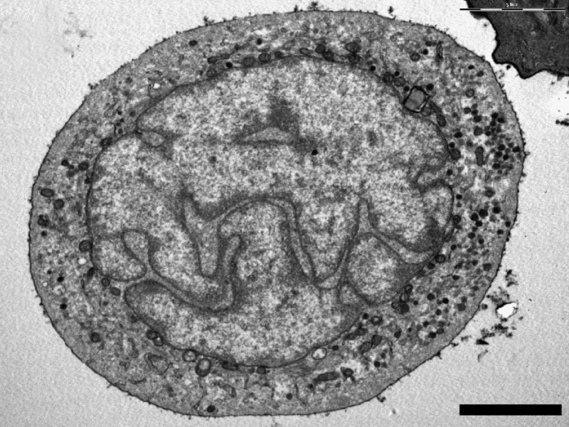
Electron microscopy analysis confirmed the identity of Merkel cells
Merkel cell (Merkel-Ranvier cell) integumentary (skin) receptor cell connected with somatosensory afferents.
- Ultrastructural analyses were carried out on cells from the enriched (Merkel cells) MC fraction.
- Up to half of the cells presented features characteristic of MCs:
- a polylobulated nucleus
- numerous typical dense-core granules in a clear cytoplasm.
Scale bar 5 µm (Stain - Osmium)
Reference
Boulais N, Pereira U, Lebonvallet N, Gobin E, Dorange G, Rougier N, Chesne C & Misery L. (2009). Merkel cells as putative regulatory cells in skin disorders: an in vitro study. PLoS ONE , 4, e6528. PMID: 19668696 DOI.
Copyright
© 2009 Boulais et al. This is an open-access article distributed under the terms of the Creative Commons Attribution License, which permits unrestricted use, distribution, and reproduction in any medium, provided the original author and source are credited.
Original file name: Figure 3. (panel a cropped from full figure, label removed and resized)
Cite this page: Hill, M.A. (2024, May 15) Embryology Merkel cell EM 01.jpg. Retrieved from https://embryology.med.unsw.edu.au/embryology/index.php/File:Merkel_cell_EM_01.jpg
- © Dr Mark Hill 2024, UNSW Embryology ISBN: 978 0 7334 2609 4 - UNSW CRICOS Provider Code No. 00098G
File history
Click on a date/time to view the file as it appeared at that time.
| Date/Time | Thumbnail | Dimensions | User | Comment | |
|---|---|---|---|---|---|
| current | 17:10, 6 June 2011 | | 984 × 738 (209 KB) | S8600021 (talk | contribs) | ==Electron microscopy analysis confirmed the identity of Merkel cells== Ultrastructural analyses were carried out on cells from the enriched MC fraction. (a, b) Up to half of the cells presented features characteristic of MCs: a polylobulated nucleus wit |
You cannot overwrite this file.
File usage
The following page uses this file: