File:Melanoblast migration.png
Melanoblast_migration.png (600 × 210 pixels, file size: 40 KB, MIME type: image/png)
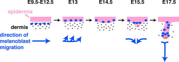
Figure 2. Schematic Depiction of Directions of Melanoblast Migration in Embryonic Mouse Skin from E9.5 to E17.5
Journal.pbio.0030372.g002.png
Pink, epithelium; black dots, dermal fibroblasts; blue ovals, melanoblasts; red dots, dermal condensate/DP.
http://www.plosbiology.org/article/info:doi/10.1371/journal.pbio.0030372
Citation: Millar SE (2005) An Ideal Society? Neighbors of Diverse Origins Interact to Create and Maintain Complex Mini-Organs in the Skin. PLoS Biol 3(11): e372. doi:10.1371/journal.pbio.0030372
Published: November 15, 2005
Copyright: © 2005 Sarah E. Millar. This is an open-access article distributed under the terms of the Creative Commons Attribution License, which permits unrestricted use, distribution, and reproduction in any medium, provided the original author and source are credited.
File history
Click on a date/time to view the file as it appeared at that time.
| Date/Time | Thumbnail | Dimensions | User | Comment | |
|---|---|---|---|---|---|
| current | 08:17, 29 September 2009 | 600 × 210 (40 KB) | S8600021 (talk | contribs) | Figure 2. Schematic Depiction of Directions of Melanoblast Migration in Embryonic Mouse Skin from E9.5 to E17.5 Journal.pbio.0030372.g002.png Pink, epithelium; black dots, dermal fibroblasts; blue ovals, melanoblasts; red dots, dermal condensate/DP. |
You cannot overwrite this file.