File:Mann1927 fig01.jpg
Original file (870 × 1,104 pixels, file size: 200 KB, MIME type: image/jpeg)
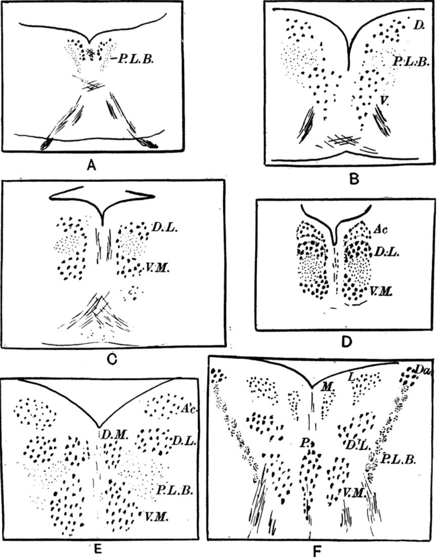
Fig. 1 A. The third nerve nucleus in a Selachian. P.L.B = posberior longitudinal bundle.
B. The nucleus in Ganoids and Teleosteans. D = dorsa.l nucleus; V = ventrs.l nucleus.
C. The nucleus in the alligator. D.L = dorso-lateral and V.M = ventro-median group of cells.
D. The nucleus in the lizard Varaxnus. Ac =accessory nucleus.
E. The nucleus in the hen. Ac = accessory nucleus; D.M = dorso-median nucleus; D.L = dorso-lateral nucleus; P.L.B : posterior longitudinal bundle; V. M = ventro-median nucleus.
F. The nucleus in man. Daznucleus of Darkschewitsch; M = medial part and L = lateral part of accessory nucleus; D.L = dorso-lateral main nucleus; V.M = ventro-median main nucleus; P = Perlia’s or median nucleus.
Reference
Mann IC. The developing third nerve nucleus in human embryos (1927) J Anat. 61(4): 424-438. PubMed 17104156
Cite this page: Hill, M.A. (2024, April 28) Embryology Mann1927 fig01.jpg. Retrieved from https://embryology.med.unsw.edu.au/embryology/index.php/File:Mann1927_fig01.jpg
- © Dr Mark Hill 2024, UNSW Embryology ISBN: 978 0 7334 2609 4 - UNSW CRICOS Provider Code No. 00098G
File history
Click on a date/time to view the file as it appeared at that time.
| Date/Time | Thumbnail | Dimensions | User | Comment | |
|---|---|---|---|---|---|
| current | 09:50, 24 February 2018 | | 870 × 1,104 (200 KB) | Z8600021 (talk | contribs) | '''Fig. 1''' A. The third nerve nucleus in a Selachian. P.L.B = posberior longitudinal bundle. B. The nucleus in Ganoids and Teleosteans. D = dorsa.l nucleus; V = ventrs.l nucleus. C. The nucleus in the alligator. D.L = dorso-lateral and V.M = ventr... |
You cannot overwrite this file.
File usage
The following page uses this file: