File:Mall1897 fig46.jpg

Original file (1,000 × 1,250 pixels, file size: 235 KB, MIME type: image/jpeg)
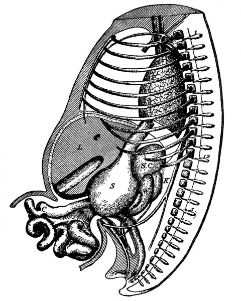
Fig. 46. Reconstruction of Embryo No. VI
Enlarged 8 times. 1-12, dorsal ganglia; S C, suprarenal capsule ; K, kidney ; W, Wolfian body ; S, stomach; C, caecum; L, liver.
The dotted area on the ventral side of the liver indicates the extent of the ventral rnesentery.
Fig. 46 (embryo VI) shows that all the tissues are becoming more definitely outlined, and the whole structure is firmer than in embryo X. The organs of the abdomen are more firmly clustered together, and the intestine has become more convoluted. The lung is much larger, and the pleural cavity extends to the ventral wall of the embryo, obscuring wholly the outline of the heart. In general it confirms everything given in Fig. 45.
Reference
Mall FP. Development of the human coelom. (1897) J Morphol. 12: 395-453.
Cite this page: Hill, M.A. (2024, April 27) Embryology Mall1897 fig46.jpg. Retrieved from https://embryology.med.unsw.edu.au/embryology/index.php/File:Mall1897_fig46.jpg
- © Dr Mark Hill 2024, UNSW Embryology ISBN: 978 0 7334 2609 4 - UNSW CRICOS Provider Code No. 00098G
File history
Click on a date/time to view the file as it appeared at that time.
| Date/Time | Thumbnail | Dimensions | User | Comment | |
|---|---|---|---|---|---|
| current | 15:47, 12 September 2017 |  | 1,000 × 1,250 (235 KB) | Z8600021 (talk | contribs) | |
| 15:45, 12 September 2017 |  | 1,245 × 1,476 (342 KB) | Z8600021 (talk | contribs) |
You cannot overwrite this file.
File usage
The following page uses this file: