File:Macklin-plate01.jpg

Original file (2,331 × 3,061 pixels, file size: 1.13 MB, MIME type: image/jpeg)
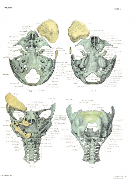
Plate 1. The skull of a human fetus of 43 millimeters greatest length
By Charles C. Macklin. (5 plates containing 47 figures)
All drawings were made by Mr. James F. Didusch according to geometric projection. With the exception of figure 7, which was made from a profile reconstruction, all figures were drawn from the original plaster-of-paris models made from human fetus No. 886 of the collection of the Carnegie Laboratory of Embiyology. The number of the model from which each figure was drawn is given, together with the magnification.
Note - the magnifications refer to teh original print versions, not the online images.
Fig. 1. Chondrocranium from above with frontal and parietal bones on right side. The densest part of the frontal bone is inclosed by a dotted hne. The basal plate is not quite horizontal, the cranial end being a little the closer to the eye of the observer. Model 1. X6.25.
Fig, 2. Chrondrocranium from below with cartilaginous branchial arch skeleton extirpated. Frontal and parietal bones are shown on right side. The basal plate is not quite horizontal, the caudal end being a httle the closer to the eye of the observer. The view is directly into the anterior nares. Model 1. X6.25.
Fig. 3. Skull from front, showing membrane bones on right side. Face is seen in frank view. The cervical vertebrae and cartilaginous branchial arch skeleton are also seen. Model 1. X6.25.
Fig. 4. Skull from back, giving a frank view of the foramen oocipitale magnum. The cervical vertebrtae are seen, their arches beiag as yet unclosed dorsaUy. Note the alignment of the hemiarch tips with the dorsal foraminal prominences, representing the extremities of the hemiarches of the occipital vertebra. The right half of the interparietal bone is seen. Model 1. X6.25.
File history
Click on a date/time to view the file as it appeared at that time.
| Date/Time | Thumbnail | Dimensions | User | Comment | |
|---|---|---|---|---|---|
| current | 16:36, 23 April 2014 | | 2,331 × 3,061 (1.13 MB) | Z8600021 (talk | contribs) | |
| 10:18, 16 February 2011 | 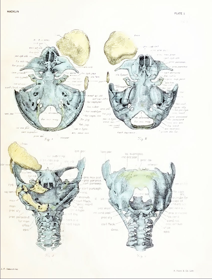 | 847 × 1,113 (182 KB) | S8600021 (talk | contribs) | ==Plate 1. The skull of a human fetus of 43 millimeters greatest length== By Charles C. Macklin. (5 plates containing 47 figures) All drawings were made by Mr. James F. Didusch according to geometric projection. With the exception of figure 7, which was |
You cannot overwrite this file.
File usage
The following 4 pages use this file: