File:MRI placenta uterus posterior upper segment.jpg
From Embryology
Size of this preview: 800 × 468 pixels. Other resolution: 1,200 × 702 pixels.
Original file (1,200 × 702 pixels, file size: 93 KB, MIME type: image/jpeg)
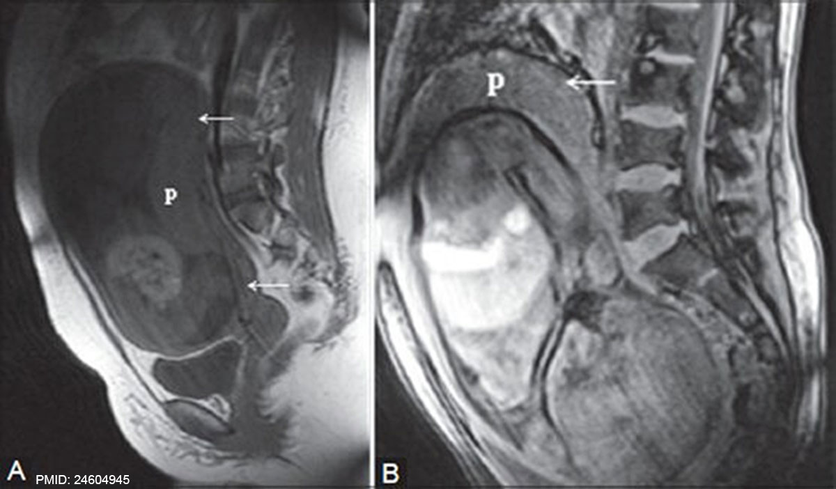
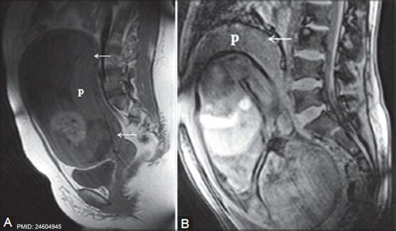
MRI placenta uterus posterior upper segment.
T1 WI showing normal placentation posteriorly at the upper segment of uterus. Both placenta and myometrium show homogeneous intermediate signal intensity.
|
|
Reference
<pubmed>24604945</pubmed>PMC3932583 | Indian J Radiol Imaging.
Varghese B, Singh N, George RA, Gilvaz S. Magnetic resonance imaging of placenta accreta. Indian J Radiol Imaging [serial online] 2013 [cited 2014 Mar 18];23:379-85. Available from: http://www.ijri.org/text.asp?2013/23/4/379/125592
Copyright
© 2007 - 2014 Indian Journal of Radiology and Imaging
File history
Click on a date/time to view the file as it appeared at that time.
| Date/Time | Thumbnail | Dimensions | User | Comment | |
|---|---|---|---|---|---|
| current | 15:30, 6 June 2014 | | 1,200 × 702 (93 KB) | Z8600021 (talk | contribs) | ==MRI placenta uterus posterior upper segment.== :'''Links:''' Placenta Development | Magnetic Resonance Imaging ===Reference=== <pubmed>24604945</pubmed>[http://www.ncbi.nlm.nih.gov/pmc/articles/PMC3932583 PMC3932583] | [http://www.ijri.org/... |
You cannot overwrite this file.
File usage
There are no pages that use this file.