File:Lung alveoli development cartoon.jpg: Difference between revisions
mNo edit summary |
m (→Reference) |
||
| Line 6: | Line 6: | ||
===Reference=== | ===Reference=== | ||
{{#pmid:6495241}} | |||
Image modified from: Scanning electron microscope study of the development of the human respiratory acinus. | Image modified from: Scanning electron microscope study of the development of the human respiratory acinus. | ||
| Line 13: | Line 13: | ||
Copyright © 1984 BMJ Publishing Group Ltd & British Thoracic Society. | Copyright © 1984 BMJ Publishing Group Ltd & British Thoracic Society. | ||
{{Footer}} | |||
[[Category:Respiratory]] [[Category:Human]][[Category:Cartoon]] | [[Category:Respiratory]] [[Category:Human]][[Category:Cartoon]] | ||
Latest revision as of 14:45, 26 May 2019
Lung Alveoli Development
Cartoon showing the stages in development of human lung alveoli.
| Lung Stage | Human | Features | Vascular | |
|---|---|---|---|---|
| Embryonic | week 4 to 5 | lung buds originate as an outgrowth from the ventral wall of the foregut where lobar division occurs | extra pulmonary artery then lobular artery | |
| Pseudoglandular | week 5 to 17 | conducting epithelial tubes surrounded by thick mesenchyme are formed, extensive airway branching | Pre-acinar arteries | |
| Canalicular | week 16 to 25 | bronchioles are produced, increasing number of capillaries in close contact with cuboidal epithelium and the beginning of alveolar epithelium development | Intra-acinar arteries | |
| Saccular | week 24 to 40 | alveolar ducts and air sacs are developed | alveolar duct arteries | |
| Alveolar | late fetal to 8 years | secondary septation occurs, marked increase of the number and size of capillaries and alveoli | alveolar capillaries | |
| embryonic stage - pseudoglandular stage - canalicular stage - saccular stage - alveolar stage Links: Species Stage Comparison | respiratory | ||||
Reference
Dilly SA. (1984). Scanning electron microscope study of the development of the human respiratory acinus. Thorax , 39, 733-42. PMID: 6495241
Image modified from: Scanning electron microscope study of the development of the human respiratory acinus.
Thorax 1984;39:733-742; doi:10.1136/thx.39.10.733 Copyright © 1984 BMJ Publishing Group Ltd & British Thoracic Society.
Cite this page: Hill, M.A. (2024, May 2) Embryology Lung alveoli development cartoon.jpg. Retrieved from https://embryology.med.unsw.edu.au/embryology/index.php/File:Lung_alveoli_development_cartoon.jpg
- © Dr Mark Hill 2024, UNSW Embryology ISBN: 978 0 7334 2609 4 - UNSW CRICOS Provider Code No. 00098G
File history
Click on a date/time to view the file as it appeared at that time.
| Date/Time | Thumbnail | Dimensions | User | Comment | |
|---|---|---|---|---|---|
| current | 14:10, 24 August 2009 | 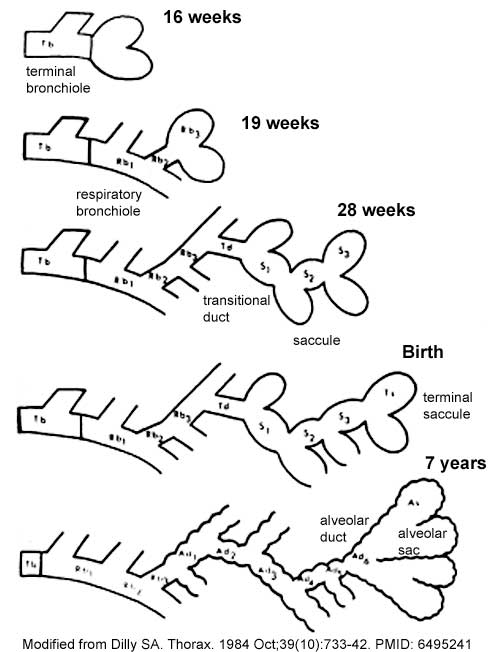 | 500 × 652 (38 KB) | MarkHill (talk | contribs) | Lung alveoli development Image modified from: Scanning electron microscope study of the development of the human respiratory acinus. Dilly SA. Thorax. 1984 Oct;39(10):733-42. [http://www.ncbi.nlm.nih.gov/pubmed/6495241 PMID: 6495241] Thorax 1984;39:733 |
You cannot overwrite this file.
File usage
The following 16 pages use this file:
- 2009 Lecture 10
- 2010 BGD Practical 12 - Second Trimester
- 2010 Lecture 10
- 2011 Lab 12 - Second Trimester
- 2011 Lab 5 - Fetal
- ANAT2341 Lab 11 - Second Trimester
- ANAT2341 Lab 12 - Second Trimester
- ANAT2341 Lab 5 - Fetal
- BGDA Practical 12 - Second Trimester
- Draft 2016
- Lecture - Fetal Development
- Lecture - Respiratory Development
- Respiratory System Development
- SH Lecture - Respiratory System Development
- Talk:2011 Lab 5 - Early Embryo
- User:Z5019799