File:Low1909 plate01.jpg

Original file (3,574 × 2,331 pixels, file size: 749 KB, MIME type: image/jpeg)
Plate I
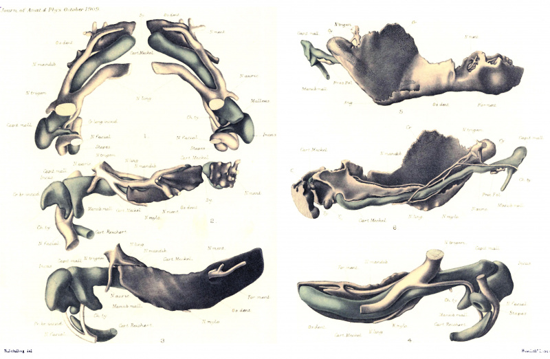
| Figs. 1 and 2. Plate-model of mandible of human embryo 18 mm. in length (Keibel’s Collection, No. 1421). The original model is 50 times enlarged, and fig. 1 represents the model as seen from above and fig. 2 as seen from the right - both drawings reduced about one-fourth.
|
Abbreviations
C. unpaired cartilaginous nodule behind symphysis. Cu. accessory cartilaginous nodule. Capit. mall. capitulum mallei. Cart. Meckel. Meckel’s cartilage. Cart. Reichert. Reichert’s cartilage. Ch. ty. N. chorda tympani. Cr. br. incud. crus brevis incudis. Cr. long. incud. crus longum incudis. Cr. coronoid accessory cartilage. Cy. condylar accessory cartilage. For. ment. foramen mentale. Manub. mall. manubrium mallei. N. auric. N. auriculo-temporalis. N. facial. N. facialis. N. mandib. N. mandibularis. N. ling. N. lingualis. N. ment. N. mentalis. IV. mylo. N. mylo~hyoideus. N. trigem. N. trigeminus. Us. dent. Dentale. Pros. Fol. processus Folianus (antr.). Sy. symphysis. |
| Historic Disclaimer - information about historic embryology pages |
|---|
 |
Reference
Low A. Further observations on the ossification of the human lower jaw. (1909) J Anat Physiol. 44(1): 83–95. PMID 17232830
Cite this page: Hill, M.A. (2024, April 27) Embryology Low1909 plate01.jpg. Retrieved from https://embryology.med.unsw.edu.au/embryology/index.php/File:Low1909_plate01.jpg
- © Dr Mark Hill 2024, UNSW Embryology ISBN: 978 0 7334 2609 4 - UNSW CRICOS Provider Code No. 00098G
File history
Click on a date/time to view the file as it appeared at that time.
| Date/Time | Thumbnail | Dimensions | User | Comment | |
|---|---|---|---|---|---|
| current | 10:57, 6 January 2017 | | 3,574 × 2,331 (749 KB) | Z8600021 (talk | contribs) | ==Plate I== Figs. 1 and 2. Plate-model of mandible of human embryo 18 mm. in length (Keibel’s Collection, No. 1421). The original model is 50 times enlarged, and fig. 1 represents the model as seen from above and fig. 2 as seen from the right - both... |
You cannot overwrite this file.
File usage
The following 2 pages use this file: