File:Lisser1911 fig11.jpg: Difference between revisions
From Embryology
mNo edit summary |
mNo edit summary |
||
| Line 2: | Line 2: | ||
[[:Category:Carnegie Embryo 43|'''Embryo no. 43''']] (16 mm.), m. constr., middle constrictor muscle; inf. constr., inferior constrictor muscle. | [[:Category:Carnegie Embryo 43|'''Embryo no. 43''']] (16 mm.), m. constr., middle constrictor muscle; inf. constr., inferior constrictor muscle. | ||
[[:Category:Carnegie Embryo 43|'''Embryo no. 43''']] - [[Carnegie stage 19|19]] [[Week 7]] | |||
{{Lisser1911 figures}} | |||
[[Category:Carnegie Embryo 43]] | |||
[[Category:Carnegie Stage 19]] | |||
[[Category:Week 7]] | |||
Latest revision as of 15:39, 12 June 2016
Fig. 11 Graphic reconstructions of pharyngeal constrictors in human Embryo 43
Embryo no. 43 (16 mm.), m. constr., middle constrictor muscle; inf. constr., inferior constrictor muscle.
- Links: fig 1 | fig 2 | fig 3 | fig 4 | fig 5 | fig 6 | fig 7 | fig 8 | fig 9 | fig 10 | fig 11 | fig 12 | fig 13 | fig 14 | Lisser 1911
Reference
Lisser H. Studies on the development of the human larynx. (1911) Amer. J Anat. 12: 27-66.
Cite this page: Hill, M.A. (2024, May 21) Embryology Lisser1911 fig11.jpg. Retrieved from https://embryology.med.unsw.edu.au/embryology/index.php/File:Lisser1911_fig11.jpg
- © Dr Mark Hill 2024, UNSW Embryology ISBN: 978 0 7334 2609 4 - UNSW CRICOS Provider Code No. 00098G
File history
Click on a date/time to view the file as it appeared at that time.
| Date/Time | Thumbnail | Dimensions | User | Comment | |
|---|---|---|---|---|---|
| current | 15:21, 12 June 2016 | 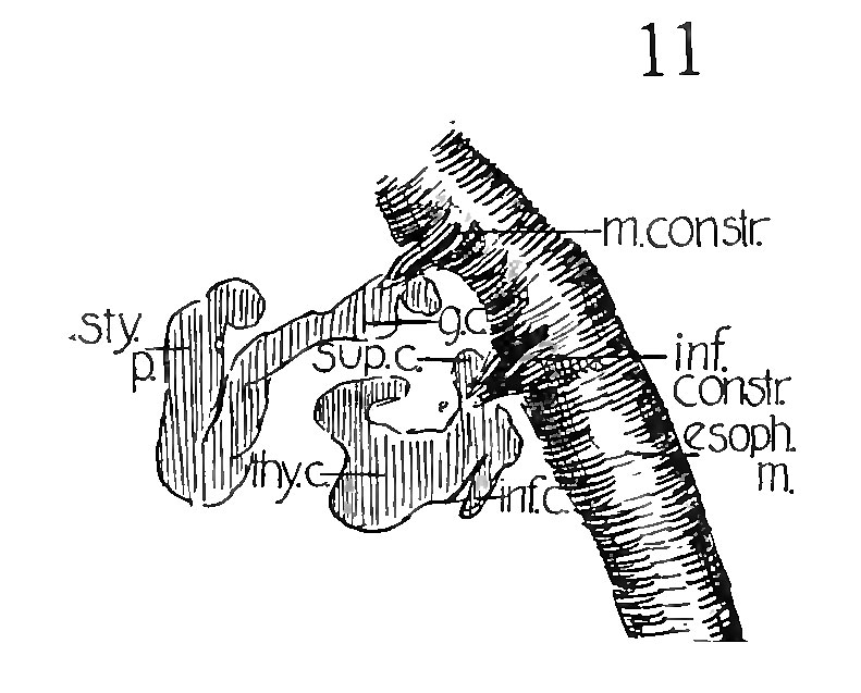 | 791 × 637 (89 KB) | Z8600021 (talk | contribs) |
You cannot overwrite this file.
File usage
The following 3 pages use this file: