File:Licata1954 fig05.jpg: Difference between revisions
From Embryology
No edit summary |
mNo edit summary |
||
| (2 intermediate revisions by the same user not shown) | |||
| Line 1: | Line 1: | ||
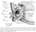
==Fig. 5 Dextral view of heart of a 25 mm embryo== | |||
With the right atrium opened, and the right venous valve cut away. The tag of tissue shown attached to septum primum is a. result of irregular resorption in the formation of ostium II. | |||
(Reconstruction X 100, illustration X 40.) | |||
{{Licata1954 figures}} | |||
===Reference=== | |||
{{Ref-Licata1954}} | |||
{{Footer}} | |||
[[Category:Heart]][[Category:Week 8]] | |||
Latest revision as of 11:15, 5 March 2017
Fig. 5 Dextral view of heart of a 25 mm embryo
With the right atrium opened, and the right venous valve cut away. The tag of tissue shown attached to septum primum is a. result of irregular resorption in the formation of ostium II.
(Reconstruction X 100, illustration X 40.)
- Links: fig 1 | fig 2 | fig 3 | fig 4 | fig 5 | fig 6 | fig 7 | fig 8 | fig 9 | fig 10 | fig 11 | fig 12 | fig 13 | fig 14 | fig 15 | fig 16 | fig 16a | fig 16b | fig 16c | fig 16d | 1954 Licata | Historic Papers | Heart Development
Reference
Licata RH. The human embryonic heart in the ninth week. (1954) Amer. J Anat., 94: 73-125. PMID 13124266
Cite this page: Hill, M.A. (2024, May 7) Embryology Licata1954 fig05.jpg. Retrieved from https://embryology.med.unsw.edu.au/embryology/index.php/File:Licata1954_fig05.jpg
- © Dr Mark Hill 2024, UNSW Embryology ISBN: 978 0 7334 2609 4 - UNSW CRICOS Provider Code No. 00098G
File history
Click on a date/time to view the file as it appeared at that time.
| Date/Time | Thumbnail | Dimensions | User | Comment | |
|---|---|---|---|---|---|
| current | 11:14, 5 March 2017 |  | 1,000 × 760 (100 KB) | Z8600021 (talk | contribs) | |
| 11:13, 5 March 2017 |  | 1,323 × 1,170 (220 KB) | Z8600021 (talk | contribs) |
You cannot overwrite this file.
File usage
The following 4 pages use this file: