File:Licata1954 fig01.jpg: Difference between revisions
From Embryology
mNo edit summary |
(Z8600021 uploaded a new version of File:Licata1954 fig01.jpg) |
(No difference)
| |
Revision as of 08:24, 5 March 2017
Fig. 1 Ventrocephalic aspect of heart of 31.5 mm embryo
Showing the superficial cardiac vessels. The arch of the aorta has been represented as if pulled cranial a little, to show the ductus arteriosus more clearly.
Reconstruction X 100, original paper illustration X 34.
Reference
Licata RH. The human embryonic heart in the ninth week. (1954) Amer. J Anat., 94: 73-125. PMID 13124266
Cite this page: Hill, M.A. (2024, May 24) Embryology Licata1954 fig01.jpg. Retrieved from https://embryology.med.unsw.edu.au/embryology/index.php/File:Licata1954_fig01.jpg
- © Dr Mark Hill 2024, UNSW Embryology ISBN: 978 0 7334 2609 4 - UNSW CRICOS Provider Code No. 00098G
File history
Click on a date/time to view the file as it appeared at that time.
| Date/Time | Thumbnail | Dimensions | User | Comment | |
|---|---|---|---|---|---|
| current | 08:24, 5 March 2017 |  | 1,000 × 725 (90 KB) | Z8600021 (talk | contribs) | |
| 08:21, 5 March 2017 | 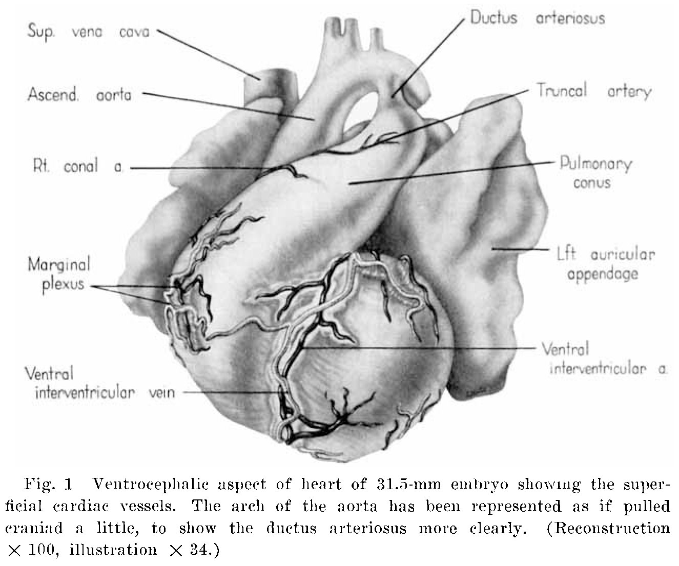 | 1,325 × 1,105 (178 KB) | Z8600021 (talk | contribs) | ===Reference=== {{Ref-Licata1954}} |
You cannot overwrite this file.
File usage
The following 4 pages use this file: