File:LattaTollman1937 fig02.jpg
LattaTollman1937_fig02.jpg (800 × 581 pixels, file size: 96 KB, MIME type: image/jpeg)
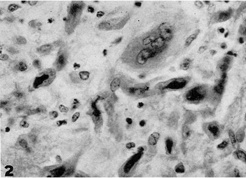
Fig. 2. A portion of the maternal mucosa lying immediately outside the implantation cavity
Showing invasion by deeply staining strands of syncytial trophoblast, the nuclei of which are mostly quite pycnotic. One large trophoblastic mass is shown which contains several typical nuclei. There are no indications of any histolytic activity on the part of this trophoblast. Note evidences of degeneration of connective tissue cells and polymorphonuelear infiltration. X 600.
- Links: fig 1 | fig 2 | fig 3 | fig 4 | fig 5 | fig 6 | fig 7 | fig 8 | fig 9 | fig 10 | fig 11 | fig 12 | fig 13 | fig 14 | fig 15 | plate 1 | plate 2 | plate 3 | 1937 Latta Tollman | Historic Papers
Reference
Latta JS. and Tollman JP. An early stage of human implantation. (1937) Anat. Rec. 69(4): 443-463.
Cite this page: Hill, M.A. (2024, April 27) Embryology LattaTollman1937 fig02.jpg. Retrieved from https://embryology.med.unsw.edu.au/embryology/index.php/File:LattaTollman1937_fig02.jpg
- © Dr Mark Hill 2024, UNSW Embryology ISBN: 978 0 7334 2609 4 - UNSW CRICOS Provider Code No. 00098G
File history
Click on a date/time to view the file as it appeared at that time.
| Date/Time | Thumbnail | Dimensions | User | Comment | |
|---|---|---|---|---|---|
| current | 12:21, 26 February 2017 |  | 800 × 581 (96 KB) | Z8600021 (talk | contribs) |
You cannot overwrite this file.
File usage
The following page uses this file:
