File:Kollmann651.jpg
Kollmann651.jpg (489 × 391 pixels, file size: 27 KB, MIME type: image/jpeg)
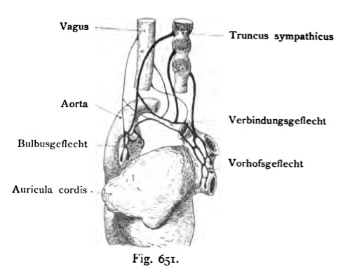
Fig. 651. Heart innervation of human embryos 10 to 19 mm CRL
Schematically. (After His Younger)
The heart of the vagus nerve are thin, thick, those of the sympathetic out- coated. By the vagus nerve and sympathetic ganglia of the next Branches form three plexuses: the trunk between the aorta and Bulbusgefl6cht Pulmonary artery, the network connection in the concavity of the aortic arch and plexus in the atrium of the upper wall of the sinus venosus and the right Forecourt. These braids form with nerve cells, which in the course of the Branches find themselves, the heart of the nervous system. Later penetrate the ends of the Nerve branches, which contain ganglion cells, with their processes in the Cardiac muscle one.
- This text is a Google translate computer generated translation and may contain many errors.
Images from - Atlas of the Development of Man (Volume 2)
(Handatlas der entwicklungsgeschichte des menschen)
- Kollmann Atlas 2: Gastrointestinal | Respiratory | Urogenital | Cardiovascular | Neural | Integumentary | Smell | Vision | Hearing | Kollmann Atlas 1 | Kollmann Atlas 2 | Julius Kollmann
- Links: Julius Kollman | Atlas Vol.1 | Atlas Vol.2 | Embryology History
| Historic Disclaimer - information about historic embryology pages |
|---|
| Pages where the terms "Historic" (textbooks, papers, people, recommendations) appear on this site, and sections within pages where this disclaimer appears, indicate that the content and scientific understanding are specific to the time of publication. This means that while some scientific descriptions are still accurate, the terminology and interpretation of the developmental mechanisms reflect the understanding at the time of original publication and those of the preceding periods, these terms, interpretations and recommendations may not reflect our current scientific understanding. (More? Embryology History | Historic Embryology Papers) |
Reference
Kollmann JKE. Atlas of the Development of Man (Handatlas der entwicklungsgeschichte des menschen). (1907) Vol.1 and Vol. 2. Jena, Gustav Fischer. (1898).
Cite this page: Hill, M.A. (2024, April 27) Embryology Kollmann651.jpg. Retrieved from https://embryology.med.unsw.edu.au/embryology/index.php/File:Kollmann651.jpg
- © Dr Mark Hill 2024, UNSW Embryology ISBN: 978 0 7334 2609 4 - UNSW CRICOS Provider Code No. 00098G
Fig. 651. Herzgeflecht menschlicher Embryonen zwischen 10 und 19 mm
Nackensteifllänge.
Schematisch. (Nach His d. J.)
Die Herznerven des Vagus sind dünn, jene des Sympathicus dick ausge- zogen. Die vom Nervus vagus und von den sympathischen Ganglien kommenden Äste bilden drei Geflechte: das Bulbusgefl6cht zwischen Truncus aortae und Pulmonalis, das Verbindungsgeflecht in der Konkavität des Aortenbogens und das Vorhofgeflecht in der oberen Wand des Sinus venosus und im rechten Vorhof. Diese Geflechte bilden mit den Nervenzellen, welche im Verlauf der Äste sich vorfinden, das Herznervensystem. Später dringen die Enden der Nervenzweige, welche Ganglienzellen enthalten, mit ihren Fortsätzen in den Herzmuskel ein.
File history
Click on a date/time to view the file as it appeared at that time.
| Date/Time | Thumbnail | Dimensions | User | Comment | |
|---|---|---|---|---|---|
| current | 09:49, 21 October 2011 |  | 489 × 391 (27 KB) | S8600021 (talk | contribs) | {{Kollmann1907}} Category:Neural Fig. 651. Herzgeflecht menschlicher Embryonen zwischen 10 und 19 mm Nackensteifllänge. Schematisch. (Nach His d. J.) Die Herznerven des Vagus sind dünn, jene des Sympathicus dick ausge- zogen. Die vom Ner |
You cannot overwrite this file.
File usage
The following page uses this file:

