File:Kollmann647.jpg

Original file (897 × 926 pixels, file size: 188 KB, MIME type: image/jpeg)
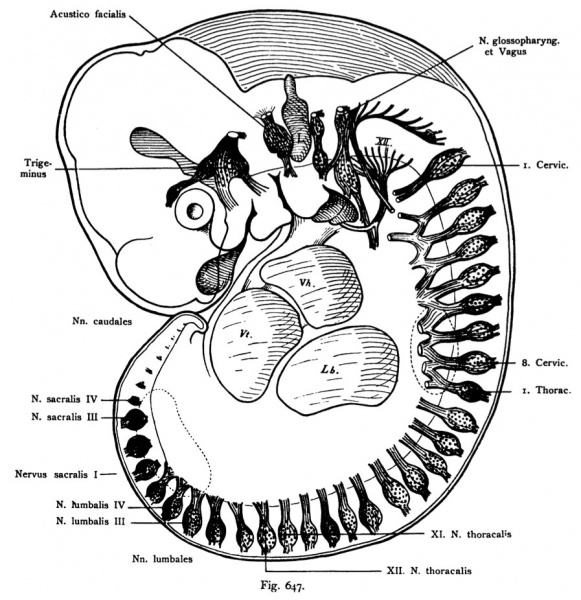
Fig. 647. Nervous System of a human embryo of 6.9 mm CRL
Seen from the left side. Magnification relative to the original about 20 times.
(After His.)
The embryo shows four pharyngeal arches. On the spokes of a wheel-together writhing bodies are seen as the main reference points for the head- the eye, the middle cranial beams and the maze bubbles L;: tonic
Xll means the hypoglossal nerve;
Vh. = Atrium of the heart;
Vt. = Ventricle of the heart;
Lb = liver. The metamerism of the peripheral nervous system is obvious in the first Line on the fuselage, but also the segmentation of the cranial nerves is well marked. The peripheral nerves are everywhere where there is no cut surfaces are specified, if drawn, as they were detectable. The Wurzelplexus plexus cervical sup. et are inferior in the making. The Sow in several places there.
- This text is a Google translate computer generated translation and may contain many errors.
Images from - Atlas of the Development of Man (Volume 2)
(Handatlas der entwicklungsgeschichte des menschen)
- Kollmann Atlas 2: Gastrointestinal | Respiratory | Urogenital | Cardiovascular | Neural | Integumentary | Smell | Vision | Hearing | Kollmann Atlas 1 | Kollmann Atlas 2 | Julius Kollmann
- Links: Julius Kollman | Atlas Vol.1 | Atlas Vol.2 | Embryology History
| Historic Disclaimer - information about historic embryology pages |
|---|
| Pages where the terms "Historic" (textbooks, papers, people, recommendations) appear on this site, and sections within pages where this disclaimer appears, indicate that the content and scientific understanding are specific to the time of publication. This means that while some scientific descriptions are still accurate, the terminology and interpretation of the developmental mechanisms reflect the understanding at the time of original publication and those of the preceding periods, these terms, interpretations and recommendations may not reflect our current scientific understanding. (More? Embryology History | Historic Embryology Papers) |
Reference
Kollmann JKE. Atlas of the Development of Man (Handatlas der entwicklungsgeschichte des menschen). (1907) Vol.1 and Vol. 2. Jena, Gustav Fischer. (1898).
Cite this page: Hill, M.A. (2024, April 27) Embryology Kollmann647.jpg. Retrieved from https://embryology.med.unsw.edu.au/embryology/index.php/File:Kollmann647.jpg
- © Dr Mark Hill 2024, UNSW Embryology ISBN: 978 0 7334 2609 4 - UNSW CRICOS Provider Code No. 00098G
Fig. 647. Nerveosystem eines menschlichen Embryo von 6,9 mm Nacken-
steifilänge,
von der linken Seite gesehen. Vergrößerung auf das Original bezogen etwa 20 mal.
(Nach His.)
Der Embryo zeigt vier Kiemenbogen. An dem radförmig zusammenge- krümmten Körper sind erkennbar als Hauptorientierungspunkte für die Kopf- nerven: das Auge, der mittlere Schädelbalken und das Labyrinthbläschen L;
Xll bedeutet den Nervus hypoglossus;
Vh. = Vorhof des Herzens ;
Vt. = Ventrikel des Herzens ;
Lb. = Leber. Die Metamerie des peripheren Nervensystems ist unverkennbar in erster Linie am Rumpf, aber auch die Metamerie der Kopfnerven tritt deutlich hervor. Die peripheren Nerven sind überall da, wo keine Schnittflächen angegeben sind, soweit gezeichnet, als sie nachweisbar waren. Die Wurzelplexus des Plexus cervicalis sup. et inferior sind im Entstehen. Die Ansäe an mehreren Stellen vorhanden.
File history
Click on a date/time to view the file as it appeared at that time.
| Date/Time | Thumbnail | Dimensions | User | Comment | |
|---|---|---|---|---|---|
| current | 09:42, 21 October 2011 |  | 897 × 926 (188 KB) | S8600021 (talk | contribs) | {{Kollmann1907}} Category:Neural Category:Week 5 Category:Spinal cord |
You cannot overwrite this file.
File usage
The following 2 pages use this file:
