File:Kollmann642.jpg
Original file (785 × 1,000 pixels, file size: 123 KB, MIME type: image/jpeg)
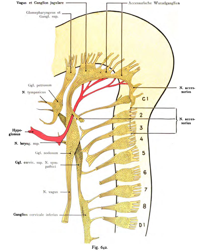
Fig. 642. Cervical and the first thoracic nerve human embryo 6th Week (17.5 mm long)
Magnified 16 times (After Streeter.)
- This text is a Google translate computer generated translation and may contain many errors.
Images from - Atlas of the Development of Man (Volume 2)
(Handatlas der entwicklungsgeschichte des menschen)
- Kollmann Atlas 2: Gastrointestinal | Respiratory | Urogenital | Cardiovascular | Neural | Integumentary | Smell | Vision | Hearing | Kollmann Atlas 1 | Kollmann Atlas 2 | Julius Kollmann
- Links: Julius Kollman | Atlas Vol.1 | Atlas Vol.2 | Embryology History
| Historic Disclaimer - information about historic embryology pages |
|---|
 |
Reference
Kollmann JKE. Atlas of the Development of Man (Handatlas der entwicklungsgeschichte des menschen). (1907) Vol.1 and Vol. 2. Jena, Gustav Fischer. (1898).
Cite this page: Hill, M.A. (2024, April 28) Embryology Kollmann642.jpg. Retrieved from https://embryology.med.unsw.edu.au/embryology/index.php/File:Kollmann642.jpg
- © Dr Mark Hill 2024, UNSW Embryology ISBN: 978 0 7334 2609 4 - UNSW CRICOS Provider Code No. 00098G
Fig. 642. Urspruog der Nerveo der Vagusgruppe, des Cervikal- und des ersten Brustnerven, ferner des Ganglion nervi sympatliici superius und inferius.
Rekonstruktion nach einem menschlichen Embryo der 6. Woche (17,5 mm lang).
16 mal vergr. (Nach Streeter.)
Die Ganglienmassen der peripheren Nerven sind heller punktiert, als die- jenigen des Sympathicus. Wie der Glossopharyngeus so hat auch der Vagus viele Wurzeln. Beim Sympathicus sind Verbindungen mit den Spinalnerven vorhanden. Das Ganglion nodosum vagi hängt mit dem Ganglion cervicale superius sympathici zusammen. Der motorische Hypoglossus ist rot. Die motorischen Wurzeln der Cervikalnerven sind durch die sensibeln Nerven größtenteils verdeckt.
File history
Click on a date/time to view the file as it appeared at that time.
| Date/Time | Thumbnail | Dimensions | User | Comment | |
|---|---|---|---|---|---|
| current | 09:32, 21 October 2011 | | 785 × 1,000 (123 KB) | S8600021 (talk | contribs) | ==Fig. 642. Cervical and the first thoracic nerve human embryo 6th Week (17.5 mm long)== Magnified 16 times (After Streeter.) {{Kollmann1907}} Category:Neural Category:Week 6 Fig. 642. Urspruog der Nerveo der Vagusgruppe, des Cervikal- und des |
You cannot overwrite this file.
File usage
The following page uses this file: