File:Kollmann639.jpg
Original file (1,068 × 655 pixels, file size: 99 KB, MIME type: image/jpeg)
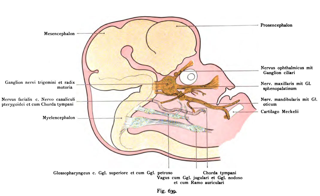
Fig. 639. The Qanslion semilunare human embryo of 15.5 mm CRL (7 Weeks old)
(Qasseri) UoD
from the main stems of the nerve ganglion hervorgelienden
in a human embryo of 15.5 mm Scheitelsteißlänge (barely seven Weeks old), seen from inside. The brain is shown in full screen for
better orientation. (According to Dixon.)
Upper and lower jaw and mouth opening after sagittal sections are connected in a way the bulbus oculi, but is again in full for better orientation Rounding drawn. Moreover, the whole face of preferred and enlarged to make room for the individual parts. There are visible:
First the ophthalmic nerve, ciliary ganglion to the top of the bulb.
Second The maxillary nerve to the ganglion. nerve and the sphenopalatine pterygoid canal (Vidii) to the geniculate ganglion of the N. facialis, which still not enclosed by the cartilaginous ear capsule.
Third The mandibular nerve with the ganglion. otic and its connection with the N. vagus (Gangl. nodosum).
4th The motor root of the trigeminal ganglion along the draws.
5th N. glossopharyngeal and vagus uppermost in their course. (About other branches, which develops in human embryos of this age already are, see Dixon.)
- This text is a Google translate computer generated translation and may contain many errors.
Images from - Atlas of the Development of Man (Volume 2)
(Handatlas der entwicklungsgeschichte des menschen)
- Kollmann Atlas 2: Gastrointestinal | Respiratory | Urogenital | Cardiovascular | Neural | Integumentary | Smell | Vision | Hearing | Kollmann Atlas 1 | Kollmann Atlas 2 | Julius Kollmann
- Links: Julius Kollman | Atlas Vol.1 | Atlas Vol.2 | Embryology History
| Historic Disclaimer - information about historic embryology pages |
|---|
 |
Reference
Kollmann JKE. Atlas of the Development of Man (Handatlas der entwicklungsgeschichte des menschen). (1907) Vol.1 and Vol. 2. Jena, Gustav Fischer. (1898).
Cite this page: Hill, M.A. (2024, April 28) Embryology Kollmann639.jpg. Retrieved from https://embryology.med.unsw.edu.au/embryology/index.php/File:Kollmann639.jpg
- © Dr Mark Hill 2024, UNSW Embryology ISBN: 978 0 7334 2609 4 - UNSW CRICOS Provider Code No. 00098G
Fig. 639. Das Qanslion semilunare (Qasseri) uod die Hauptstämme der aus
dem Ganglion hervorgelienden Nerven
bei einem menschlichen Embryo von 15,5 mm Scheitelsteißlänge (kaum sieben Wochen alt), von innen gesehen. Das Gehirn ist im Vollbild eingezeichnet zur
besseren Orientierung. (Nach Dixon.)
Ober- und Unterkiefer und Mundspalte sind nach Sagittalschnitten kon- struiert, der Bulbus oculi ist aber wieder zur besseren Orientierung in voller Rundung eingezeichnet. Überdies ist der ganze Gesichtsteil vorgezogen und vergrößert, um Raum für die einzelnen Teile zu gewinnen. Es sind sichtbar gemacht :
1. der Nervus ophthalmicus mit dem Ganglion ciliare oberhalb des Bulbus.
2. Der Nervus maxillaris mit dem Gangl. sphenopalatinum und dem Nervus canalis pterygoidei (Vidii) zum Ganglion geniculi des N. facialis, der noch nicht von der knorpeligen Ohrkapsel umschlossen ist.
3. Der Nervus mandibularis mit dem Gangl. oticum und dessen Verbindung mit dem N. vagus (Gangl. nodosum).
4. Die motorische Wurzel des Trigeminus zieht dem Ganglion entlang.
5. N. glossopharyngeus und Vagus in ihrem obersten Verlauf. (Über andere Äste, die bei menschlichen Embryonen dieses Alters schon entwickelt sind, siehe Dixon.)
File history
Click on a date/time to view the file as it appeared at that time.
| Date/Time | Thumbnail | Dimensions | User | Comment | |
|---|---|---|---|---|---|
| current | 09:28, 21 October 2011 | | 1,068 × 655 (99 KB) | S8600021 (talk | contribs) | ==Fig. 639. The Qanslion semilunare human embryo of 15.5 mm CRL (7 Weeks old)== (Qasseri) UoD from the main stems of the nerve ganglion hervorgelienden in a human embryo of 15.5 mm Scheitelsteißlänge (barely seven Weeks old), seen from inside. The b |
You cannot overwrite this file.
File usage
The following page uses this file: