File:Kollmann638.jpg
Kollmann638.jpg (667 × 467 pixels, file size: 42 KB, MIME type: image/jpeg)
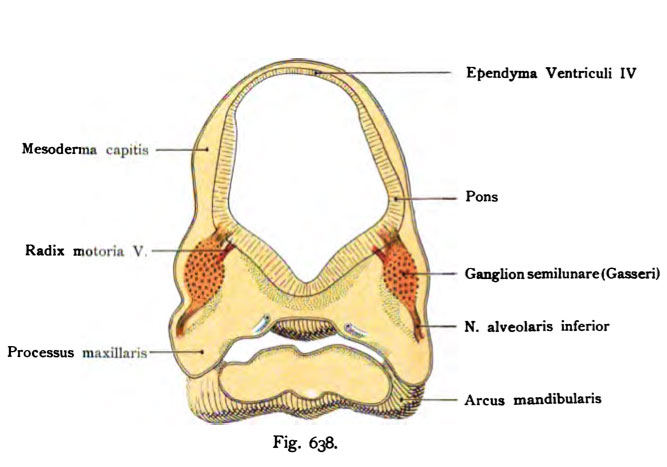
Fig. 638. Semilunar ganglion human embryo of 6.9 mm length (4 weeks old)
(Qasseri).
Horizontal section through the head of a human embryo of 6.9 mm length (4 weeks old).
Reconstruction. (According to Dixon.)
On the average, the brain appears in the tube brocken bow, with the beginning of the myelencephalon is made. The semilunar ganglion (Gasserian) comes with its sensory and motor root in the mesoderm of the Head out. The outgoing from the ganglion nerve is the nerve alveolar inferiorly. The reconstruction, however, the course of the nerves in the lower jaw not taken. Ciliary ganglion, the sphenopalatine ganglion and the otic are not yet detectable. Between the maxillary and mandibular protrusion the primitive oral cavity visible.
- This text is a Google translate computer generated translation and may contain many errors.
Images from - Atlas of the Development of Man (Volume 2)
(Handatlas der entwicklungsgeschichte des menschen)
- Kollmann Atlas 2: Gastrointestinal | Respiratory | Urogenital | Cardiovascular | Neural | Integumentary | Smell | Vision | Hearing | Kollmann Atlas 1 | Kollmann Atlas 2 | Julius Kollmann
- Links: Julius Kollman | Atlas Vol.1 | Atlas Vol.2 | Embryology History
| Historic Disclaimer - information about historic embryology pages |
|---|
| Pages where the terms "Historic" (textbooks, papers, people, recommendations) appear on this site, and sections within pages where this disclaimer appears, indicate that the content and scientific understanding are specific to the time of publication. This means that while some scientific descriptions are still accurate, the terminology and interpretation of the developmental mechanisms reflect the understanding at the time of original publication and those of the preceding periods, these terms, interpretations and recommendations may not reflect our current scientific understanding. (More? Embryology History | Historic Embryology Papers) |
Reference
Kollmann JKE. Atlas of the Development of Man (Handatlas der entwicklungsgeschichte des menschen). (1907) Vol.1 and Vol. 2. Jena, Gustav Fischer. (1898).
Cite this page: Hill, M.A. (2024, April 28) Embryology Kollmann638.jpg. Retrieved from https://embryology.med.unsw.edu.au/embryology/index.php/File:Kollmann638.jpg
- © Dr Mark Hill 2024, UNSW Embryology ISBN: 978 0 7334 2609 4 - UNSW CRICOS Provider Code No. 00098G
Fig. 638. Ganglion semilunare (Qasseri).
Horizontalschnitt durch den Kopf eines menschlichen Embryo von 6,9 mm Länge
(4 Wochen alt).
Rekonstruktion. (Nach Dixon.)
Auf dem Schnitt erscheint das Hirnrohr im Bereich der BrOckenbeuge, wobei der Anfang des Myelencephalon getroffen ist. Das Ganglion semilunare (Gasseri) mit seiner sensibeln und motorischen Wurzel tritt im Mesoderm des Kopfes hervor. Der aus dem Ganglion abgehende Nerv ist der N. alveolaris inferior. Die Rekonstruktion hat aber den Verlauf des Nerven im Unterkiefer nicht getroffen. Ganglion ciliare, Ganglion sphenopalatinum und oticum sind noch nicht nachweisbar. Zwischen Oberkieferfortsatz und Mandibularbogen ist die primitive Mundhöhle sichtbar.
File history
Click on a date/time to view the file as it appeared at that time.
| Date/Time | Thumbnail | Dimensions | User | Comment | |
|---|---|---|---|---|---|
| current | 09:26, 21 October 2011 |  | 667 × 467 (42 KB) | S8600021 (talk | contribs) | ==Fig. 638. Semilunar ganglion human embryo of 6.9 mm length (4 weeks old)== (Qasseri). Horizontal section through the head of a human embryo of 6.9 mm length (4 weeks old). Reconstruction. (According to Dixon.) On the average, the brain appears in th |
You cannot overwrite this file.
File usage
The following page uses this file:

