File:Kollmann636.jpg

Original file (759 × 756 pixels, file size: 65 KB, MIME type: image/jpeg)
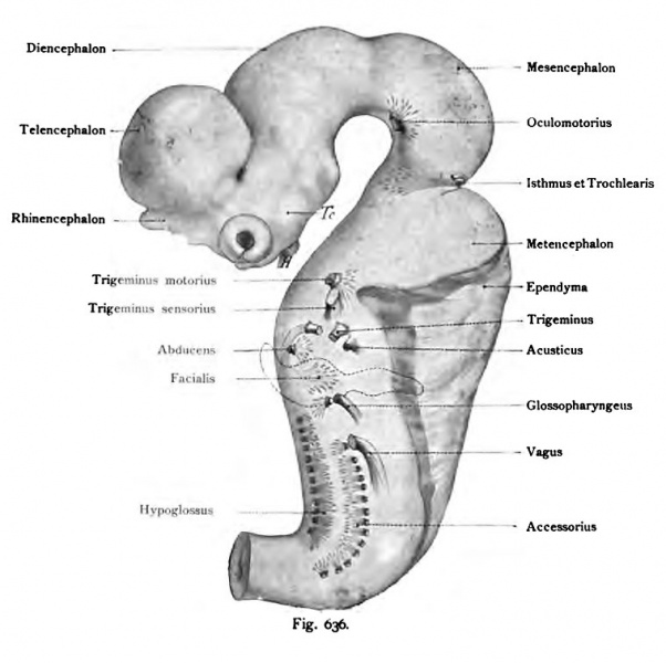
The exits of the cranial nerves in the brain of a human embryo of 10.2 mm CRL
About i8 times magnified. (Reconstruction by His.)
The hypoglossal follows the paths of the motor spinal nerves. It emerges at the base. This body meets later the lateral sulcus anterior to the medulla oblongata. The glossopharyngeal, vagus and accessory embryonic brain rely on its lateral surface, along the edge, which forms the base of the wing panel, later called the sulcus lateralis posterior. The above-mentioned nerves exit points along the form enclosed in the embryonic neural tube Nervenkemen an almost regular series. When the spinal accessory nerve and H3rpoglossus the number of exit points is calculated only approximately. Next orally and laterally from the maze of bubbles occurs abducens nerve and N. produce facial, and dorsal to it by the auditory nerve. The type of the spinal nerves still follows the oculomotor nerve with respect to the exit point.
- This text is a Google translate computer generated translation and may contain many errors.
Images from - Atlas of the Development of Man (Volume 2)
(Handatlas der entwicklungsgeschichte des menschen)
- Kollmann Atlas 2: Gastrointestinal | Respiratory | Urogenital | Cardiovascular | Neural | Integumentary | Smell | Vision | Hearing | Kollmann Atlas 1 | Kollmann Atlas 2 | Julius Kollmann
- Links: Julius Kollman | Atlas Vol.1 | Atlas Vol.2 | Embryology History
| Historic Disclaimer - information about historic embryology pages |
|---|
| Pages where the terms "Historic" (textbooks, papers, people, recommendations) appear on this site, and sections within pages where this disclaimer appears, indicate that the content and scientific understanding are specific to the time of publication. This means that while some scientific descriptions are still accurate, the terminology and interpretation of the developmental mechanisms reflect the understanding at the time of original publication and those of the preceding periods, these terms, interpretations and recommendations may not reflect our current scientific understanding. (More? Embryology History | Historic Embryology Papers) |
Reference
Kollmann JKE. Atlas of the Development of Man (Handatlas der entwicklungsgeschichte des menschen). (1907) Vol.1 and Vol. 2. Jena, Gustav Fischer. (1898).
Cite this page: Hill, M.A. (2024, April 27) Embryology Kollmann636.jpg. Retrieved from https://embryology.med.unsw.edu.au/embryology/index.php/File:Kollmann636.jpg
- © Dr Mark Hill 2024, UNSW Embryology ISBN: 978 0 7334 2609 4 - UNSW CRICOS Provider Code No. 00098G
Fig. 636. Die Austrittsstellen der Hirnnerven an dem Gehirn eines menschlichen Embryo von 10,2 mm Nackensteifilänge.
Etwa i8 mal vergrößert. (Rekonstruktion von His.)
Der Hypoglossus folgt dem T3T)us der motorischen Spinal-Nerven. Er tritt an der Grundplatte hervor. Diese Stelle entspricht später dem Sulcus lateralis anterior der Medulla oblongata. Der Nervus glossopharyngeus, vagus und accessorius verlassen das embryonale Hirn an seiner lateralen Oberfläche, längs der Kante, welche die Grundplatte mit der Flügelplatte bildet, später Sulcus lateralis posterior genannt. Die eben erwähnten Nervenaustrittsstellen bilden samt den in dem embryonalen Nervenrohr eingeschlossenen Nervenkemen eine fast regelmäßige Reihe. Bei dem Nervus accessorius und H3rpoglossus ist die Zahl der Austrittsstellen nur annähernd angegeben. Weiter oral und lateral, vor dem Labyrinthbläschen tritt der Nervus abducens und N. facialis hervor, und dorsal von ihm der Acusticus. Dem Typus der Spinalnerven folgt noch der Oculomotorius bezüglich der Austrittsstelle.
File history
Click on a date/time to view the file as it appeared at that time.
| Date/Time | Thumbnail | Dimensions | User | Comment | |
|---|---|---|---|---|---|
| current | 17:26, 17 October 2011 |  | 759 × 756 (65 KB) | S8600021 (talk | contribs) | {{Kollmann1907}} Category:Human Category:Fetal Category:Neural Fig. 636. Die Austrittsstellen der Hirnnerven an dem Gehirn eines mensch- lichen Embryo von 10,2 mm Nackensteifilänge. Etwa i8 mal vergrößert. (Rekonstruktion von His.) |
You cannot overwrite this file.
File usage
The following 2 pages use this file:
