File:Kollmann630.jpg
Kollmann630.jpg (412 × 322 pixels, file size: 17 KB, MIME type: image/jpeg)
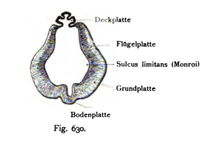
Fig. 630. Section through a human embryo 5 weeks
Magnified 22 times
(after His.)
The repetitive structure of the walls of the brain tube is here again and especially clearly noticeable: the base plate, base plate, the alar plate and the cover plate. At the boundary between primary and wing plate runs the sulcus limitans (Fig. 602 and 604). The top plate is a Ependymlamelle, emerges from the case of mammals and humans, the pineal Corpus. The sulcus limitans is identical to the sulcus limitans (Monro) the systematic anatomy, which is continued in the hypothalamic sulcus.
- This text is a Google translate computer generated translation and may contain many errors.
Images from - Atlas of the Development of Man (Volume 2)
(Handatlas der entwicklungsgeschichte des menschen)
- Kollmann Atlas 2: Gastrointestinal | Respiratory | Urogenital | Cardiovascular | Neural | Integumentary | Smell | Vision | Hearing | Kollmann Atlas 1 | Kollmann Atlas 2 | Julius Kollmann
- Links: Julius Kollman | Atlas Vol.1 | Atlas Vol.2 | Embryology History
| Historic Disclaimer - information about historic embryology pages |
|---|
 |
Reference
Kollmann JKE. Atlas of the Development of Man (Handatlas der entwicklungsgeschichte des menschen). (1907) Vol.1 and Vol. 2. Jena, Gustav Fischer. (1898).
Cite this page: Hill, M.A. (2024, April 28) Embryology Kollmann630.jpg. Retrieved from https://embryology.med.unsw.edu.au/embryology/index.php/File:Kollmann630.jpg
- © Dr Mark Hill 2024, UNSW Embryology ISBN: 978 0 7334 2609 4 - UNSW CRICOS Provider Code No. 00098G
Fig. 630. Schnitt durch das Zwischenhu-n eines menschlichen Embryo
von 5 Wochen.
22 mal vergr. (Nach His.)
Die immer wiederkehrende Gliederung der Wandungen des Hirnrohres ist hier aufs Neue und besonders übersichtlich bemerkbar: die Bodenplatte, die Grundplatte, die Flügelplatte und die Deckplatte. An der Grenze zwischen Grund- und Flügelplatte verläuft der Sulcus limitans (Fig. 602 und 604). Die Deckplatte stellt eine Ependymlamelle dar, aus der bei den Säugetieren und dem Menschen das Corpus pineale hervorgeht. Der Sulcus limitans ist identisch mit dem Sulcus limitans (Monroi) der systematischen Anatomie, der sich in den Sulcus hypothalamicus fortsetzt.
File history
Click on a date/time to view the file as it appeared at that time.
| Date/Time | Thumbnail | Dimensions | User | Comment | |
|---|---|---|---|---|---|
| current | 17:22, 17 October 2011 | | 412 × 322 (17 KB) | S8600021 (talk | contribs) | {{Kollmann1907}} Category:Human Category:Fetal Category:Neural Fig. 630. Schnitt durch das Zwischenhu-n eines menschlichen Embryo von 5 Wochen. 22 mal vergr. (Nach His.) Die immer wiederkehrende Gliederung der Wandungen des Hirnro |
You cannot overwrite this file.
File usage
The following page uses this file: