File:Kollmann629.jpg
Original file (747 × 647 pixels, file size: 110 KB, MIME type: image/jpeg)
- This text is a Google translate computer generated translation and may contain many errors.
Images from - Atlas of the Development of Man (Volume 2)
(Handatlas der entwicklungsgeschichte des menschen)
- Kollmann Atlas 2: Gastrointestinal | Respiratory | Urogenital | Cardiovascular | Neural | Integumentary | Smell | Vision | Hearing | Kollmann Atlas 1 | Kollmann Atlas 2 | Julius Kollmann
- Links: Julius Kollman | Atlas Vol.1 | Atlas Vol.2 | Embryology History
| Historic Disclaimer - information about historic embryology pages |
|---|
 |
Reference
Kollmann JKE. Atlas of the Development of Man (Handatlas der entwicklungsgeschichte des menschen). (1907) Vol.1 and Vol. 2. Jena, Gustav Fischer. (1898).
Cite this page: Hill, M.A. (2024, April 28) Embryology Kollmann629.jpg. Retrieved from https://embryology.med.unsw.edu.au/embryology/index.php/File:Kollmann629.jpg
- © Dr Mark Hill 2024, UNSW Embryology ISBN: 978 0 7334 2609 4 - UNSW CRICOS Provider Code No. 00098G
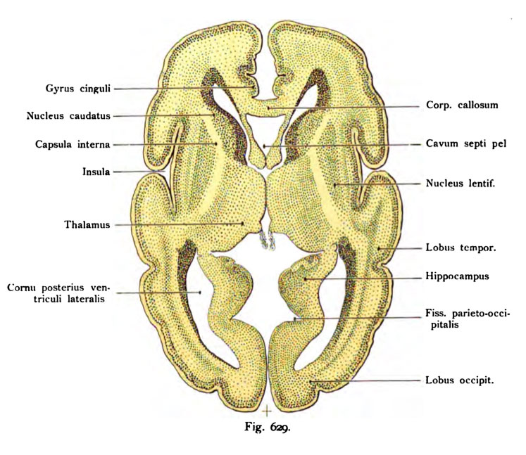
Fig. 629. Horizontalschoitt durch das Qehiro eines meoschlicheo Fetus
des 6. Monats.
(Anatomische Sammlung in Basel.)
Der Schnitt geht unter dem Corpus callosum hindurch trifft aber vorn das Genu corporis callosi, die beiden Lamellen des Septum pellucidum, die Columnae fornicis und das Foramen interventriculare (Monroi). Die Seitenven- trikel sind zweimal getroffen als Cornu anterius und als Cornu inferius. In der Mitte springen die Thalami medial hervor und ein Teil des Ventriculus tertius ist geöffnet. In der Wand des Hemisphärenhirns ist durch dunkle Schattierung der Nucleus caudatus (caput und cauda) erkennbar, die Capsula interna (Genu, pars frontalis und occipitalis) und im Cornu inferius der Ventriculi laterales der Hippocampus, die Digitationes hippocampi, die Fissura hippocampi und jene Schichten, welche auch in der Hemisphärenwand vorkommen. Von Windungen sind bemerkbar : der Gyrus cinguli, von dem Balken getrennt durch den Sulcus corporis callosi ; nach vorn ist dieser Gyrus cinguli begrenzt durch den Sulcus cinguli. Vor diesem Sulcus bis zur medialen Kante der Hemisphären und noch übergreifend auf die laterale Oberfläche erstreckt sich der Gyrus frontalis superior. Die Entwicklung der Windungen ist auf der einen Seite etwas weiter fortgeschritten als auf der anderen Seite, vorausgesetzt, daß hier nicht eine postmortale Erscheinung vorliegt.
File history
Click on a date/time to view the file as it appeared at that time.
| Date/Time | Thumbnail | Dimensions | User | Comment | |
|---|---|---|---|---|---|
| current | 17:21, 17 October 2011 | | 747 × 647 (110 KB) | S8600021 (talk | contribs) | {{Kollmann1907}} Category:Human Category:Fetal Category:Neural Fig. 629. Horizontalschoitt durch das Qehiro eines meoschlicheo Fetus des 6. Monats. (Anatomische Sammlung in Basel.) Der Schnitt geht unter dem Corpus callosum hindurch |
You cannot overwrite this file.
File usage
The following page uses this file: