File:Kollmann628.jpg
Original file (823 × 543 pixels, file size: 126 KB, MIME type: image/jpeg)
- This text is a Google translate computer generated translation and may contain many errors.
Images from - Atlas of the Development of Man (Volume 2)
(Handatlas der entwicklungsgeschichte des menschen)
- Kollmann Atlas 2: Gastrointestinal | Respiratory | Urogenital | Cardiovascular | Neural | Integumentary | Smell | Vision | Hearing | Kollmann Atlas 1 | Kollmann Atlas 2 | Julius Kollmann
- Links: Julius Kollman | Atlas Vol.1 | Atlas Vol.2 | Embryology History
| Historic Disclaimer - information about historic embryology pages |
|---|
 |
Reference
Kollmann JKE. Atlas of the Development of Man (Handatlas der entwicklungsgeschichte des menschen). (1907) Vol.1 and Vol. 2. Jena, Gustav Fischer. (1898).
Cite this page: Hill, M.A. (2024, April 28) Embryology Kollmann628.jpg. Retrieved from https://embryology.med.unsw.edu.au/embryology/index.php/File:Kollmann628.jpg
- © Dr Mark Hill 2024, UNSW Embryology ISBN: 978 0 7334 2609 4 - UNSW CRICOS Provider Code No. 00098G
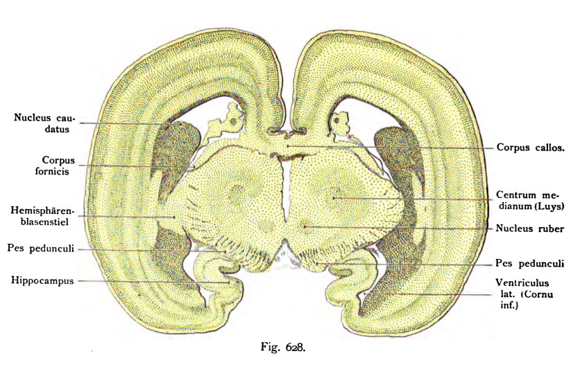
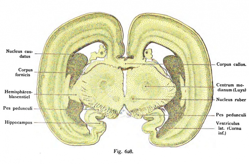
Fig. 628. Frootalschiiitt durch das Qehirn eines mensclilichen Fetus von
5 Monaten.
(Anatomische Sammlung in Basel.)
Der Schnitt trifft den Thalamus und die vordere Partie der Hirnschenkel. Die Gehirnoberfläche zeigt rechts keine Furchen, die links sichtbaren sind wohl postmortal. Zwischen den beiden Thalami klafft der Ventriculus tertius bedeckt von der Tela chorioidea media. Darüber befindet sich der Balken und zu beiden Seiten an ihn anschließend die platten GewOlbsschenkel (Corpora fomicis). Nach oben ist der Balken abgegrenzt durch den Sulcus corporis callosi. In dem Vorderhorn der Seitenventrikel ist das Corpus striatum getroffen ebenso im Unterhorn. Der Hemisphärenblasenstiel (Hochsteller , Hirnstiel His) nament- lich links deutlich, verbindet die Hemisphärenblasenwand mit dem Thalamus. Die mediale Wand des Unterhorns wird von dem Hippocampus gebildet Es ist noch keine Randfurche entwickelt.
File history
Click on a date/time to view the file as it appeared at that time.
| Date/Time | Thumbnail | Dimensions | User | Comment | |
|---|---|---|---|---|---|
| current | 17:20, 17 October 2011 | | 823 × 543 (126 KB) | S8600021 (talk | contribs) | {{Kollmann1907}} Category:Human Category:Fetal Category:Neural Fig. 628. Frootalschiiitt durch das Qehirn eines mensclilichen Fetus von 5 Monaten. (Anatomische Sammlung in Basel.) Der Schnitt trifft den Thalamus und die vordere Parti |
You cannot overwrite this file.
File usage
The following page uses this file: