File:Kollmann625.jpg
Kollmann625.jpg (733 × 454 pixels, file size: 62 KB, MIME type: image/jpeg)
- This text is a Google translate computer generated translation and may contain many errors.
Images from - Atlas of the Development of Man (Volume 2)
(Handatlas der entwicklungsgeschichte des menschen)
- Kollmann Atlas 2: Gastrointestinal | Respiratory | Urogenital | Cardiovascular | Neural | Integumentary | Smell | Vision | Hearing | Kollmann Atlas 1 | Kollmann Atlas 2 | Julius Kollmann
- Links: Julius Kollman | Atlas Vol.1 | Atlas Vol.2 | Embryology History
| Historic Disclaimer - information about historic embryology pages |
|---|
 |
Reference
Kollmann JKE. Atlas of the Development of Man (Handatlas der entwicklungsgeschichte des menschen). (1907) Vol.1 and Vol. 2. Jena, Gustav Fischer. (1898).
Cite this page: Hill, M.A. (2024, April 28) Embryology Kollmann625.jpg. Retrieved from https://embryology.med.unsw.edu.au/embryology/index.php/File:Kollmann625.jpg
- © Dr Mark Hill 2024, UNSW Embryology ISBN: 978 0 7334 2609 4 - UNSW CRICOS Provider Code No. 00098G
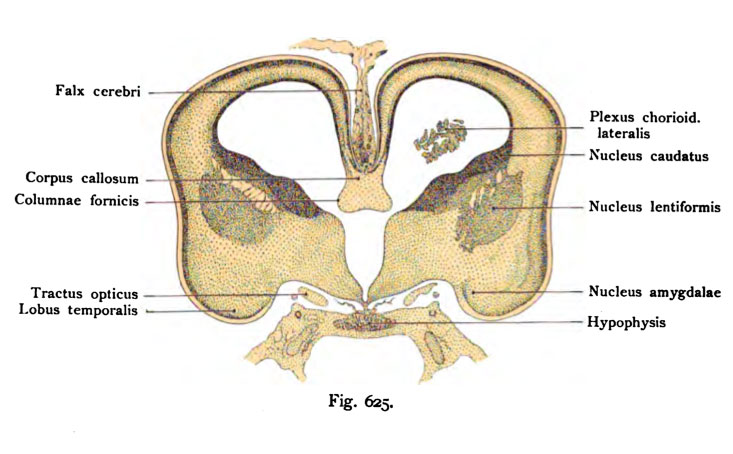
Fig. 625. Frontalschnitt durch das Hemisphärenhirn emes menschlichen Fetus
vom Ende des 4. Monats.
(8 fache Vergrößerung.) (Nach Hochstetter.)
Der Frontalschnitt trifft das Foramen Monroi und den vordersten Abschnitt der dritten Hirnkammer mit dem Infundibulum und der H3rpophysis. Die Ein- ziehung der seitlichen Hemisphärenwand entspricht der Fossa cerebri lateralis (Sylvii), die ventrale Ausladung dem späteren Vorderende des Lobus temporalis mit der Anlage des Nucleus amygdalae. Medianwärts befindet sich beiderseits der Tractus opticus. Die beiden Hemisphärenblasen stehen oberhalb des Foramen interventriculare (Monroi) durch die Lamina terminalis miteinander in Verbindung, mit einer seitlichen Verdickung, in der man die Fornixfaserung erkennt und darüber eine Faserung, welche wie in der Fig. 624 beschaffen, wahrscheinlich dem Balken angehört. Corpus striatum, innere Kapsel, Nucleus lentiformis, Schichten der Hemisphärenwand wie in Fig. 624.
File history
Click on a date/time to view the file as it appeared at that time.
| Date/Time | Thumbnail | Dimensions | User | Comment | |
|---|---|---|---|---|---|
| current | 17:18, 17 October 2011 | | 733 × 454 (62 KB) | S8600021 (talk | contribs) | {{Kollmann1907}} Category:Human Category:Fetal Category:Neural Fig. 625. Frontalschnitt durch das Hemisphärenhirn emes menschlichen Fetus vom Ende des 4. Monats. (8 fache Vergrößerung.) (Nach Hochstetter.) Der Frontalschnitt trif |
You cannot overwrite this file.
File usage
The following page uses this file: