File:Kollmann622.jpg
Original file (712 × 617 pixels, file size: 46 KB, MIME type: image/jpeg)
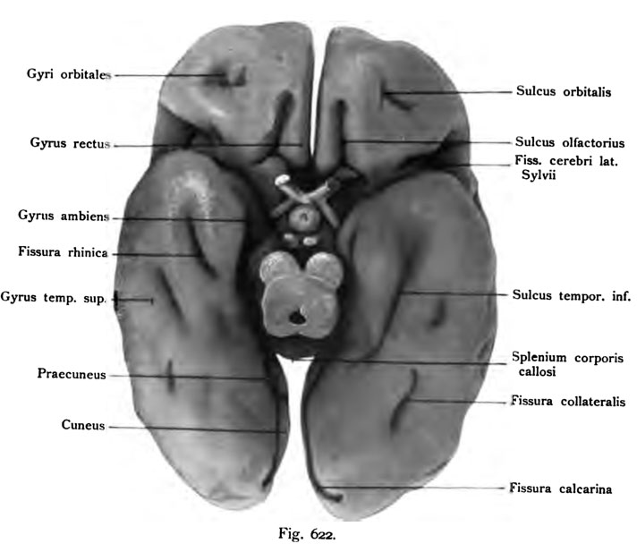
Fig. 622. fetus from the end of 7th month
(Compare to figures 620 and 621)
(Anatomical Collection in Basel)
The cerebellum, including the MeduUa oblongata, olfactory bulb and tract are also removed. The fissura cerebri lateralis (Sylvian) is deep and the entrance to it is far frontal lobe On the Sulcus olfactorius are very fully. Of the orbital sulci is only developed one. Lobe on the right temporalis sulcus temporalis inferior handsomely designed, the left is? only fragmentary case of contrast, the calcarine fissure is visible only in the shortened course, just as the parieto-occipital fissure, together with its sequel. Some links are called lobules and Gyn. The fissure rhinica can be seen here and in Fig 623 In the deep, close to the cerebral peduncles and the hippocampal fissure runs.
- This text is a Google translate computer generated translation and may contain many errors.
Images from - Atlas of the Development of Man (Volume 2)
(Handatlas der entwicklungsgeschichte des menschen)
- Kollmann Atlas 2: Gastrointestinal | Respiratory | Urogenital | Cardiovascular | Neural | Integumentary | Smell | Vision | Hearing | Kollmann Atlas 1 | Kollmann Atlas 2 | Julius Kollmann
- Links: Julius Kollman | Atlas Vol.1 | Atlas Vol.2 | Embryology History
| Historic Disclaimer - information about historic embryology pages |
|---|
 |
Reference
Kollmann JKE. Atlas of the Development of Man (Handatlas der entwicklungsgeschichte des menschen). (1907) Vol.1 and Vol. 2. Jena, Gustav Fischer. (1898).
Cite this page: Hill, M.A. (2024, April 28) Embryology Kollmann622.jpg. Retrieved from https://embryology.med.unsw.edu.au/embryology/index.php/File:Kollmann622.jpg
- © Dr Mark Hill 2024, UNSW Embryology ISBN: 978 0 7334 2609 4 - UNSW CRICOS Provider Code No. 00098G
Fig. 622. Circumvolutiooes pallii eines menschlicheo Fetus vom Ende des
7. Monats.
(Vergl. die Figuren 620 und 621.) (Anatomische Sammlung in Basel)
Das Cerebellum samt der MeduUa oblongata, ferner Bulbus und Tractus olfactorius sind entfernt. Die Fissura cerebri lateralis (Sylvii) ist tief und der Eingang *zu ihr weit Am Lobus frontalis ist der Sulcus olfactorius sehr voll- kommen. Von den Sulci orbitales ist nur einer entwickelt. Am Lobus tempo- ralis ist rechts der Sulcus temporalis inferior ansehnlich ausgebildet, links ist die? erst bruchstückweise der Fall Dagegen ist die Fissura calcarina, freilich nur in der Verkürzung sichtbar, ebenso die Fissura parieto-occipitalis samt ihrer Fortsetzung. Links sind einige Lobuli und Gyn bezeichnet. Die Fissura rhinica ist hier und in der Fig. 623 zu sehen. In der Tiefe, dicht neben den Pedunculi cerebri verläuft die Fissura hippocampi.
File history
Click on a date/time to view the file as it appeared at that time.
| Date/Time | Thumbnail | Dimensions | User | Comment | |
|---|---|---|---|---|---|
| current | 17:16, 17 October 2011 | | 712 × 617 (46 KB) | S8600021 (talk | contribs) | {{Kollmann1907}} Category:Human Category:Fetal Category:Neural Fig. 622. Circumvolutiooes pallii eines menschlicheo Fetus vom Ende des 7. Monats. (Vergl. die Figuren 620 und 621.) (Anatomische Sammlung in Basel) Das Cerebellum samt |
You cannot overwrite this file.
File usage
The following page uses this file: