File:Kollmann621.jpg
Original file (976 × 550 pixels, file size: 49 KB, MIME type: image/jpeg)
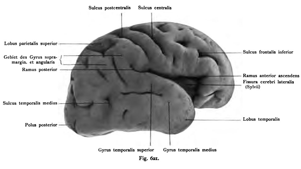
Fig. 621. Right hemisphere human Fetus from the end of the 7th month
Viewed from the side. (See Fig. 620) (Anatomical Collection in Basel)
The lateral fissure (of Sylvius) is wide open and reveals the depth of the island. Developed Deudich the ramus posterior lateral fissure. On the frontal lobe of the inferior frontal sulcus is deep and impressive curved, it limits the inferior frontal lobe (Sprachwindung). The sulcus centralis prae-is, as the comparison with the results Norma verticalis, separated into two separate sections created. The sulcus centralis (Rolandi) of the central sulcus and posterior to run in parallel, but terminate far above the fissura cerebri lateralis. At the parietal lobe above the interparietal sulcus is visible. After the sheath edge is toward the superior parietal lobule, below the inferior lobule parietaUs. The supramarginal gyrus angularis and are not exactly limited, nor the gyri lateral occipital. The boundary between the parietal and occipital lobes is deeply incised by the fissura parieto-occipitalis (see Fig. 623).
- This text is a Google translate computer generated translation and may contain many errors.
Images from - Atlas of the Development of Man (Volume 2)
(Handatlas der entwicklungsgeschichte des menschen)
- Kollmann Atlas 2: Gastrointestinal | Respiratory | Urogenital | Cardiovascular | Neural | Integumentary | Smell | Vision | Hearing | Kollmann Atlas 1 | Kollmann Atlas 2 | Julius Kollmann
- Links: Julius Kollman | Atlas Vol.1 | Atlas Vol.2 | Embryology History
| Historic Disclaimer - information about historic embryology pages |
|---|
 |
Reference
Kollmann JKE. Atlas of the Development of Man (Handatlas der entwicklungsgeschichte des menschen). (1907) Vol.1 and Vol. 2. Jena, Gustav Fischer. (1898).
Cite this page: Hill, M.A. (2024, April 28) Embryology Kollmann621.jpg. Retrieved from https://embryology.med.unsw.edu.au/embryology/index.php/File:Kollmann621.jpg
- © Dr Mark Hill 2024, UNSW Embryology ISBN: 978 0 7334 2609 4 - UNSW CRICOS Provider Code No. 00098G
Fig. 621. Circumvolutiones pallii einer rechten Hemisphäre. Menschlicher
Fetus vom Ende des 7. Monats.
Von der Seite gesehen. (Vergl. Fig. 620.) Vergrößert.
(Anatomische Sammlung in Basel)
Die Fissura lateralis (Sylvii) ist weit geöffnet und läßt die Tiefe der Insel erkennen. Deudich entwickelt ist der Ramus posterior fissurae lateralis. Am Lobus frontalis ist der Sulcus frontalis inferior tief und ansehnlich gekrümmt; er begrenzt den Lobulus frontalis inferior (Sprachwindung). Der Sulcus prae- centralis ist, wie die Vergleichung mit der Norma verticalis ergibt, in zwei ge- trennten Abschnitten angelegt. Der Sulcus centralis (Rolandi) und der Sulcus centralis posterior laufen parallel, endigen aber weit oberhalb der Fissura cerebri lateralis. Am Scheitellappen ist oben der Sulcus interparietalis sichtbar. Nach der Mantelkante hin befindet sich der Lobulus parietalis superior, unterhalb der Lobulus parietaUs inferior. Der Gyrus supramarginalis und angularis sind noch nicht genau begrenzt, ebensowenig die Gyri occipitales laterales. Die Grenze zwischen Parietal- und Occipitallappen ist tief eingeschnitten durch die Fissura parieto-occipitalis (vergl. die Fig. 623).
File history
Click on a date/time to view the file as it appeared at that time.
| Date/Time | Thumbnail | Dimensions | User | Comment | |
|---|---|---|---|---|---|
| current | 17:15, 17 October 2011 | | 976 × 550 (49 KB) | S8600021 (talk | contribs) | {{Kollmann1907}} Category:Human Category:Fetal Category:Neural Fig. 621. Circumvolutiones pallii einer rechten Hemisphäre. Menschlicher Fetus vom Ende des 7. Monats. Von der Seite gesehen. (Vergl. Fig. 620.) Vergrößert. (Anatomisch |
You cannot overwrite this file.
File usage
The following page uses this file: