File:Kollmann617.jpg
Kollmann617.jpg (688 × 395 pixels, file size: 41 KB, MIME type: image/jpeg)
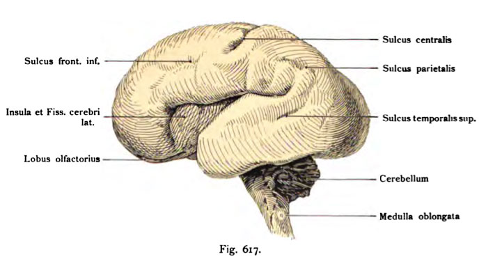
Fig. 617. 30 cm long female Fetus (End of the 5th month)
Viewed from the side. Lying inside the skull.
(Anatomical Collection in Basel)
Appearance of the sulci and thus delineation of individual lobes: lobes at a somewhat younger than Fig. 618 fetus To the most striking limitation of the island is by the fissura cerebri lateralis (Sylvian), whose anterior ascending ramus is only just visible as an incision, the posterior ramus appears very strong. The central sulcus is in its upper section develops, the pre-and postcentral sulcus barely recognizable. In contrast, the inferior frontal sulcus and the superior temporal sulcus is present in small beginnings, much less the parietal sulcus.
- This text is a Google translate computer generated translation and may contain many errors.
Images from - Atlas of the Development of Man (Volume 2)
(Handatlas der entwicklungsgeschichte des menschen)
- Kollmann Atlas 2: Gastrointestinal | Respiratory | Urogenital | Cardiovascular | Neural | Integumentary | Smell | Vision | Hearing | Kollmann Atlas 1 | Kollmann Atlas 2 | Julius Kollmann
- Links: Julius Kollman | Atlas Vol.1 | Atlas Vol.2 | Embryology History
| Historic Disclaimer - information about historic embryology pages |
|---|
| Pages where the terms "Historic" (textbooks, papers, people, recommendations) appear on this site, and sections within pages where this disclaimer appears, indicate that the content and scientific understanding are specific to the time of publication. This means that while some scientific descriptions are still accurate, the terminology and interpretation of the developmental mechanisms reflect the understanding at the time of original publication and those of the preceding periods, these terms, interpretations and recommendations may not reflect our current scientific understanding. (More? Embryology History | Historic Embryology Papers) |
Reference
Kollmann JKE. Atlas of the Development of Man (Handatlas der entwicklungsgeschichte des menschen). (1907) Vol.1 and Vol. 2. Jena, Gustav Fischer. (1898).
Cite this page: Hill, M.A. (2024, April 27) Embryology Kollmann617.jpg. Retrieved from https://embryology.med.unsw.edu.au/embryology/index.php/File:Kollmann617.jpg
- © Dr Mark Hill 2024, UNSW Embryology ISBN: 978 0 7334 2609 4 - UNSW CRICOS Provider Code No. 00098G
Fig. 617. Qeliirn eines 30 cm langen weiblichen Fetus
(Ende des 5. Monates). Von der Seite gesehen. Im Innern des Schädels liegend.
(Anatomische Sammlung in Basel)
Auftreten der Sulci und dadurch Abgrenzung einzelner Hirnlappen: Lobi bei einem etwas jüngeren Fetus als Fig. 618. Am auffallendsten ist die Um- grenzung der Insel durch die Fissura cerebri lateralis (Sylvii), deren Ramus anterior ascendens nur eben als Einschnitt erkennbar ist, der Ramus posterior erscheint dagegen sehr stark. Der Sulcus centralis ist in seinem oberen Ab- schnitt entwickelt; der Sulcus prae- und postcentralis noch kaum erkennbar. Dagegen ist der Sulcus frontalis inferior und der Sulcus temporalis superior in kleinen Anfängen vorhanden, weniger deutlich der Sulcus parietalis.
File history
Click on a date/time to view the file as it appeared at that time.
| Date/Time | Thumbnail | Dimensions | User | Comment | |
|---|---|---|---|---|---|
| current | 17:04, 17 October 2011 |  | 688 × 395 (41 KB) | S8600021 (talk | contribs) | {{Kollmann1907}} Category:Human Category:Neural Fig. 617. Qeliirn eines 30 cm langen weiblichen Fetus (Ende des 5. Monates). Von der Seite gesehen. Im Innern des Schädels liegend. (Anatomische Sammlung in Basel) Auftreten der Sulci und |
You cannot overwrite this file.
File usage
The following page uses this file:

