File:Kollmann615.jpg
Kollmann615.jpg (660 × 496 pixels, file size: 35 KB, MIME type: image/jpeg)
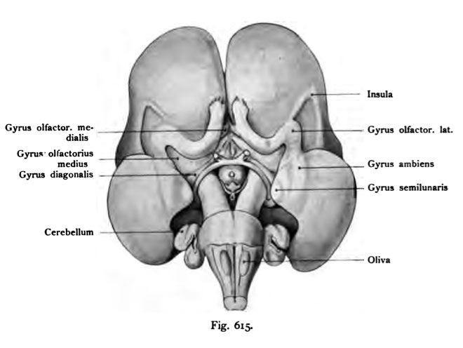
Fig. 615. Base of the brain of a human fetus from the beginning of 4th Month
As seen from below. The same brain as in Fig 613, and 614.
(Anatomical Collection in Basel)
The rhinencephalon has developed remarkably in the human fetus at this age. You can see the two olfactory Bulbis with short, almost without tract. After the rear, close to three sequels, as the gyrus olfactorius lateralis, medialis and medius be distinguished. The gyrus olf lateral moves to the medial edge of the island, but turns at an acute angle: angulus gyri olf. lateralis is out, after the peak of the temporal lobe, to move into the semilunar gyrus and the gyrus ambiens, which then result in more connections. Posteriorly from the olfactory gyrus. the curved medial lamina terminalis is visible, then follows the chiasma nervorum opti corum, just before the chiasm, the lamina terminalis with a ovalgn location of the fenestra laminae terminalis, and the nerves and optic tracts, still very small. In the midline dorsally then the tuber cinereum follows with the cloverleaf-shaped eminence saccularis after hinteh framed by the narrow and low mammillary bodies. The other details are understood by the nomenclature.
- This text is a Google translate computer generated translation and may contain many errors.
Images from - Atlas of the Development of Man (Volume 2)
(Handatlas der entwicklungsgeschichte des menschen)
- Kollmann Atlas 2: Gastrointestinal | Respiratory | Urogenital | Cardiovascular | Neural | Integumentary | Smell | Vision | Hearing | Kollmann Atlas 1 | Kollmann Atlas 2 | Julius Kollmann
- Links: Julius Kollman | Atlas Vol.1 | Atlas Vol.2 | Embryology History
| Historic Disclaimer - information about historic embryology pages |
|---|
| Pages where the terms "Historic" (textbooks, papers, people, recommendations) appear on this site, and sections within pages where this disclaimer appears, indicate that the content and scientific understanding are specific to the time of publication. This means that while some scientific descriptions are still accurate, the terminology and interpretation of the developmental mechanisms reflect the understanding at the time of original publication and those of the preceding periods, these terms, interpretations and recommendations may not reflect our current scientific understanding. (More? Embryology History | Historic Embryology Papers) |
Reference
Kollmann JKE. Atlas of the Development of Man (Handatlas der entwicklungsgeschichte des menschen). (1907) Vol.1 and Vol. 2. Jena, Gustav Fischer. (1898).
Cite this page: Hill, M.A. (2024, April 28) Embryology Kollmann615.jpg. Retrieved from https://embryology.med.unsw.edu.au/embryology/index.php/File:Kollmann615.jpg
- © Dr Mark Hill 2024, UNSW Embryology ISBN: 978 0 7334 2609 4 - UNSW CRICOS Provider Code No. 00098G
Fig. 615. Basis des Gehirns von einem menschlichen Fetus vom Anfang des
4. Monates, von unten gesehen.
Das nämliche Gehirn wie in der Fig. 613 und 614. (Anatomische Sammlung in Basel)
Das Rhinencephalon ist bei dem menschlichen Fetus dieses Alters auffallend entwickelt. Man erkennt die beiden Olfactorii mit kurzen Bulbis, fast ohne Tractus. Nach hinten schließen sich drei Fortsetzungen an, die als Gyrus olfactorius lateralis, medialis und medius unterschieden werden. Der Gyrus olf lateralis zieht an den medialen Rand der Insel, biegt aber in spitzem Winkel: Angulus gyri olf. lateralis um, geht nach der Spitze des Schläfenlappens, um in den Gyrus semilunaris und in den Gyrus ambiens überzugehen, wo sich dann weitere Verbindungen ergeben. Dorsal von den Gyrus olfact. medialis wird die gewölbte Lamina terminalis sichtbar, dann folgt das Chiasma nervorum opticorum, unmittelbar vor dem Chiasma die Lamina terminalis mit einer ovalgn Stelle, der Fenestra laminae terminalis; die Nervi und Tractus optici, noch sehr klein. In der Mittellinie dorsalwärts folgt dann das Tuber cinereum mit der kleeblattförmigen Eminentia saccularis nach hinteh umrahmt von den schmalen und niedrigen Corpora mamillaria. Die übrigen Einzelheiten sind durch die Namengebung verständlich.
File history
Click on a date/time to view the file as it appeared at that time.
| Date/Time | Thumbnail | Dimensions | User | Comment | |
|---|---|---|---|---|---|
| current | 17:03, 17 October 2011 |  | 660 × 496 (35 KB) | S8600021 (talk | contribs) | {{Kollmann1907}} Category:Human Category:Neural Fig. 615. Basis des Gehirns von einem menschlichen Fetus vom Anfang des 4. Monates, von unten gesehen. Das nämliche Gehirn wie in der Fig. 613 und 614. (Anatomische Sammlung in Basel) Da |
You cannot overwrite this file.
File usage
The following page uses this file:

