File:Kollmann610.jpg
Original file (898 × 555 pixels, file size: 51 KB, MIME type: image/jpeg)
- This text is a Google translate computer generated translation and may contain many errors.
Images from - Atlas of the Development of Man (Volume 2)
(Handatlas der entwicklungsgeschichte des menschen)
- Kollmann Atlas 2: Gastrointestinal | Respiratory | Urogenital | Cardiovascular | Neural | Integumentary | Smell | Vision | Hearing | Kollmann Atlas 1 | Kollmann Atlas 2 | Julius Kollmann
- Links: Julius Kollman | Atlas Vol.1 | Atlas Vol.2 | Embryology History
| Historic Disclaimer - information about historic embryology pages |
|---|
 |
Reference
Kollmann JKE. Atlas of the Development of Man (Handatlas der entwicklungsgeschichte des menschen). (1907) Vol.1 and Vol. 2. Jena, Gustav Fischer. (1898).
Cite this page: Hill, M.A. (2024, April 28) Embryology Kollmann610.jpg. Retrieved from https://embryology.med.unsw.edu.au/embryology/index.php/File:Kollmann610.jpg
- © Dr Mark Hill 2024, UNSW Embryology ISBN: 978 0 7334 2609 4 - UNSW CRICOS Provider Code No. 00098G
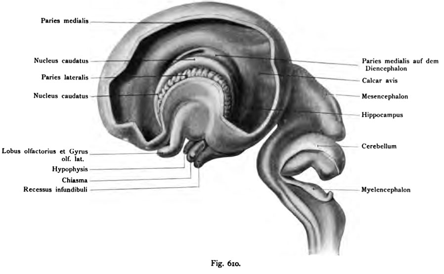
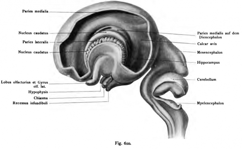
Fig. 610. Gehirn eines menschlichen Fetus vom 3. Monat und von 42 mm
Scheitelsteifilänge.
(3. Monat.) (Teilweise nach His.)
Die Hemisphärenblase bedeckt schon das ganze Zwischenhirn. Das Mittel- hirn liegt aber noch frei. Daran schließt sich die Anlage des Cerebellum und des Nach- oder Markhirns. Die linke Hemisphärenblase ist von außen her weit eröffnet, doch blieb die Insel unverletzt. Der Plexus chorioideus ist aus der Hemisphärenblase entfernt Folgende Einzelheiten sind in dem noch weiten, primitiven Seiten Ventrikel bemerkbar: die verdickte laterale Wand der Hemi- sphärenblase, der bogenförmige Nucleus caudatus, der die Fossa cerebri lateralis (Sylvii) umgreift, und sich ventral und dorsal hinabsenkt bis auf den Boden des primitiven Seiten Ventrikels. Ferner ist sichtbar : die mediale Hemisphären wand, welche das Zwischenhirn bedeckt, ferner ein Schlitz: Fissura chorioidea zum Eintritt der Art chorioidea, und bogenförmige Erhebungen im hinteren Ab- schnitt der Hemisphärenblase, welche den Calcar avis und den Hippocampus andeuten.
File history
Click on a date/time to view the file as it appeared at that time.
| Date/Time | Thumbnail | Dimensions | User | Comment | |
|---|---|---|---|---|---|
| current | 16:59, 17 October 2011 | | 898 × 555 (51 KB) | S8600021 (talk | contribs) | {{Kollmann1907}} Category:Human Category:Neural Fig. 610. Gehirn eines menschlichen Fetus vom 3. Monat und von 42 mm Scheitelsteifilänge. (3. Monat.) (Teilweise nach His.) Die Hemisphärenblase bedeckt schon das ganze Zwischenhirn. Da |
You cannot overwrite this file.
File usage
The following page uses this file: