File:Kollmann584.jpg
Kollmann584.jpg (457 × 461 pixels, file size: 26 KB, MIME type: image/jpeg)
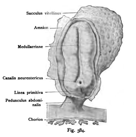
Fig. 584. The first unit of the nervous system in a human embryo
The length of the whole embryo was 2 mm.
(After Graf Spee)
The amnion, which lies across the embryo is torn, side but still partially present. On the sandal-shaped blastoderm seen in the longitudinal direction running a respectable groove that neural canal. It is bounded on both sides by two powerful, well-along different running beads, the medullary folds. These diverge in front, there which later caused the cerebral vesicles. medullary vesicles also run behind the apart, and take the neurenteric canal between himself and later even the primitive groove.
- This text is a Google translate computer generated translation and may contain many errors.
Images from - Atlas of the Development of Man (Volume 2)
(Handatlas der entwicklungsgeschichte des menschen)
- Kollmann Atlas 2: Gastrointestinal | Respiratory | Urogenital | Cardiovascular | Neural | Integumentary | Smell | Vision | Hearing | Kollmann Atlas 1 | Kollmann Atlas 2 | Julius Kollmann
- Links: Julius Kollman | Atlas Vol.1 | Atlas Vol.2 | Embryology History
| Historic Disclaimer - information about historic embryology pages |
|---|
| Pages where the terms "Historic" (textbooks, papers, people, recommendations) appear on this site, and sections within pages where this disclaimer appears, indicate that the content and scientific understanding are specific to the time of publication. This means that while some scientific descriptions are still accurate, the terminology and interpretation of the developmental mechanisms reflect the understanding at the time of original publication and those of the preceding periods, these terms, interpretations and recommendations may not reflect our current scientific understanding. (More? Embryology History | Historic Embryology Papers) |
Reference
Kollmann JKE. Atlas of the Development of Man (Handatlas der entwicklungsgeschichte des menschen). (1907) Vol.1 and Vol. 2. Jena, Gustav Fischer. (1898).
Cite this page: Hill, M.A. (2024, April 28) Embryology Kollmann584.jpg. Retrieved from https://embryology.med.unsw.edu.au/embryology/index.php/File:Kollmann584.jpg
- © Dr Mark Hill 2024, UNSW Embryology ISBN: 978 0 7334 2609 4 - UNSW CRICOS Provider Code No. 00098G
Fig. 584. Die erste Anlage des Nervensystems bei einem menschlichen Embryo.
Die Länge des ganzen Keimlings betrug 2 mm.
(Nach Graf S p e e.)
Das Amnion, das über die Embryonalanlage hinwegzieht, ist durchgerissen, seitlich aber noch teilweise vorhanden. Auf der sandalenförmigen Keimhaut zeigt sich in der Längsrichtung verlaufend eine ansehnliche Rinne, die MeduUar- rinne. Sie ist beiderseits begrenzt von zwei mächtigen, ebenfalls längs ver- laufenden Wülsten, den Medullarwülsten. Diese laufen vorn auseinander, dort wo später die Hirnblasen entstehen. Hinten laufen die MeduUarwülste ebenfalls auseinander, und nehmen den Canalis neurentericus zwischen sich und später auch noch die Primitivrinne.
File history
Click on a date/time to view the file as it appeared at that time.
| Date/Time | Thumbnail | Dimensions | User | Comment | |
|---|---|---|---|---|---|
| current | 16:24, 17 October 2011 |  | 457 × 461 (26 KB) | S8600021 (talk | contribs) | {{Kollmann1907}} |
You cannot overwrite this file.
File usage
The following page uses this file:

