File:Kollmann542.jpg
Kollmann542.jpg (732 × 520 pixels, file size: 74 KB, MIME type: image/jpeg)
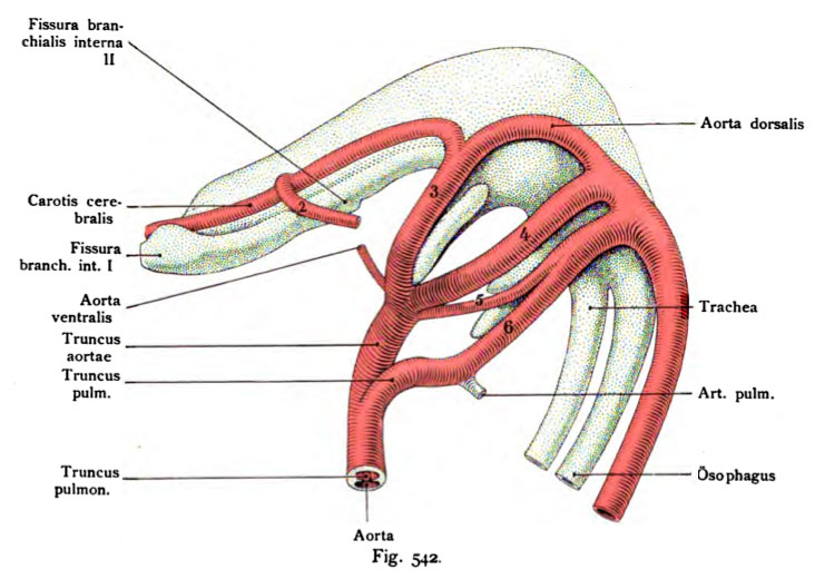
Fig. 542. Aortic arch to the left of a human embryo of 9 mm length
Original image magnification x 75 times.
(According to Tandler.)
In this embryo is the most recent discovered in the sixth time aortic
developed aortic arch, the fifth well in the series. Thus the number of aortic arch directly to the reptiles. the
Division of the bulbus aortae into a trunk of the aorta and pulmonary trunk is just going to take place from above downwards are the aortas wells are located in the foregut. The inner gill-pouches are shown particularly clearly. It should be noted that the outer contour of pharyngeal epithelium has served as a border.
- This text is a Google translate computer generated translation and may contain many errors.
Images from - Atlas of the Development of Man (Volume 2)
(Handatlas der entwicklungsgeschichte des menschen)
- Kollmann Atlas 2: Gastrointestinal | Respiratory | Urogenital | Cardiovascular | Neural | Integumentary | Smell | Vision | Hearing | Kollmann Atlas 1 | Kollmann Atlas 2 | Julius Kollmann
- Links: Julius Kollman | Atlas Vol.1 | Atlas Vol.2 | Embryology History
| Historic Disclaimer - information about historic embryology pages |
|---|
 |
Reference
Kollmann JKE. Atlas of the Development of Man (Handatlas der entwicklungsgeschichte des menschen). (1907) Vol.1 and Vol. 2. Jena, Gustav Fischer. (1898).
Cite this page: Hill, M.A. (2024, April 28) Embryology Kollmann542.jpg. Retrieved from https://embryology.med.unsw.edu.au/embryology/index.php/File:Kollmann542.jpg
- © Dr Mark Hill 2024, UNSW Embryology ISBN: 978 0 7334 2609 4 - UNSW CRICOS Provider Code No. 00098G
Fig. 542. Aortenbogen der linken Seite von einem menschlichen Embryo von
9 mm sröfiter Länge.
Vergr. 75 mal. (Nach Tandler.)
Bei diesem Embryo ist der erst in der jüngsten Zeit entdeckte 6. Aorten- bogen, der fünfte in der Reihe deutlich entwickelt. Dadurch schliefet sich der Mensch bezüglich der Zahl der Aortenbogen direkt an die Reptilien an. Die Teilung des Bulbus aortae in einen Truncus aortae und Truncus pulmonalis ist eben im Begriff, von oben nach abwärts sich zu vollziehen. Die Aorten liegen in Vertiefungen des Kopfdarms. Die inneren Kiementaschen werden dadurch besonders deutlich erkennbar. Dabei ist zu bemerken, daß der äußere Kontur des Pharynxepithels als Grenze gedient hat.
File history
Click on a date/time to view the file as it appeared at that time.
| Date/Time | Thumbnail | Dimensions | User | Comment | |
|---|---|---|---|---|---|
| current | 00:11, 17 October 2011 | | 732 × 520 (74 KB) | S8600021 (talk | contribs) | {{Kollmann1907}} |
You cannot overwrite this file.
File usage
The following page uses this file: