File:Kollmann533.jpg
Kollmann533.jpg (717 × 594 pixels, file size: 76 KB, MIME type: image/jpeg)
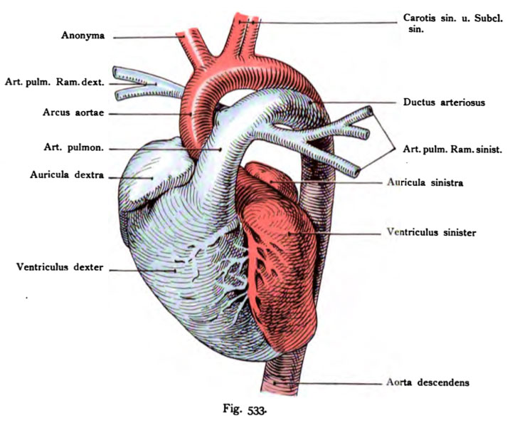
Fig. 533. Heart of a newborn viewed from the front and placed in the vertical direction
(Anatomical Collection in Basel)
The right half is blue, the left in red and the descending aorta other hand, has a mixed color from the preceding two colors to the mixture of blood is suggested through the inclusion of the ductus arteriosus.
- This text is a Google translate computer generated translation and may contain many errors.
Images from - Atlas of the Development of Man (Volume 2)
(Handatlas der entwicklungsgeschichte des menschen)
- Kollmann Atlas 2: Gastrointestinal | Respiratory | Urogenital | Cardiovascular | Neural | Integumentary | Smell | Vision | Hearing | Kollmann Atlas 1 | Kollmann Atlas 2 | Julius Kollmann
- Links: Julius Kollman | Atlas Vol.1 | Atlas Vol.2 | Embryology History
| Historic Disclaimer - information about historic embryology pages |
|---|
 |
Reference
Kollmann JKE. Atlas of the Development of Man (Handatlas der entwicklungsgeschichte des menschen). (1907) Vol.1 and Vol. 2. Jena, Gustav Fischer. (1898).
Cite this page: Hill, M.A. (2024, April 27) Embryology Kollmann533.jpg. Retrieved from https://embryology.med.unsw.edu.au/embryology/index.php/File:Kollmann533.jpg
- © Dr Mark Hill 2024, UNSW Embryology ISBN: 978 0 7334 2609 4 - UNSW CRICOS Provider Code No. 00098G
Fig. 533. Herz eines Neugeborenen, injiziert, von vorn gesehen und in die senkrechte Richtung gebracht.
(Anatomische Sammlung in Basel)
Die rechte Herzhälfte ist blau, die linke rot gefärbt, die Aorta descendens besitzt dagegen eine Mischfarbe aus den beiden vorhergehenden Farben, um die Mischung des. Blutes durch die Aufnahme des Ductus arteriosus anzudeuten.
File history
Click on a date/time to view the file as it appeared at that time.
| Date/Time | Thumbnail | Dimensions | User | Comment | |
|---|---|---|---|---|---|
| current | 00:09, 17 October 2011 | | 717 × 594 (76 KB) | S8600021 (talk | contribs) | {{Kollmann1907}} |
You cannot overwrite this file.
File usage
The following 2 pages use this file: