File:Kollmann531.jpg
Kollmann531.jpg (688 × 570 pixels, file size: 88 KB, MIME type: image/jpeg)
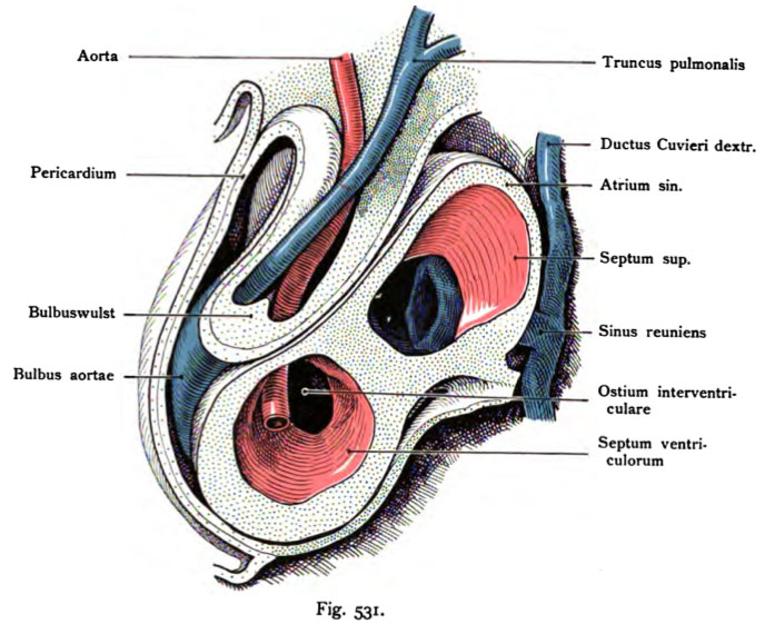
Fig. 531. Heart included in the pericardium of a human embryo of 7.5 mm body length
about 4 weeks old.
(After His.) Reconstruction.
The heart is open laterally, above the left atrium and its wide Communication with the right, in between the superior septum. in the right Atrium occurs dorsal to the sinus opening, surrounded by the right and left Sinus flap. In the section taken from the left ventricle is the septum to see the inferior and still wide communication with the right ventricle. Ventral to the above-mentioned parts of the aortic bulb is open to the to show the two blood flows in his heart: the aorta and pulmonary trunk. The currents move within (clear), endothelial, which treated here as arteries. The beginning of separation into an aorta and Pulmonary artery is carried out by the Bulbuswülste (one is visible), which emanate from opposite sides of the pipe still common.
- This text is a Google translate computer generated translation and may contain many errors.
Images from - Atlas of the Development of Man (Volume 2)
(Handatlas der entwicklungsgeschichte des menschen)
- Kollmann Atlas 2: Gastrointestinal | Respiratory | Urogenital | Cardiovascular | Neural | Integumentary | Smell | Vision | Hearing | Kollmann Atlas 1 | Kollmann Atlas 2 | Julius Kollmann
- Links: Julius Kollman | Atlas Vol.1 | Atlas Vol.2 | Embryology History
| Historic Disclaimer - information about historic embryology pages |
|---|
| Pages where the terms "Historic" (textbooks, papers, people, recommendations) appear on this site, and sections within pages where this disclaimer appears, indicate that the content and scientific understanding are specific to the time of publication. This means that while some scientific descriptions are still accurate, the terminology and interpretation of the developmental mechanisms reflect the understanding at the time of original publication and those of the preceding periods, these terms, interpretations and recommendations may not reflect our current scientific understanding. (More? Embryology History | Historic Embryology Papers) |
Reference
Kollmann JKE. Atlas of the Development of Man (Handatlas der entwicklungsgeschichte des menschen). (1907) Vol.1 and Vol. 2. Jena, Gustav Fischer. (1898).
Cite this page: Hill, M.A. (2024, April 28) Embryology Kollmann531.jpg. Retrieved from https://embryology.med.unsw.edu.au/embryology/index.php/File:Kollmann531.jpg
- © Dr Mark Hill 2024, UNSW Embryology ISBN: 978 0 7334 2609 4 - UNSW CRICOS Provider Code No. 00098G
Fig. 531. Herz im Herzbeutel eingeschlossen von einem menschlichen Embryo von 7,5 mm Körperlänge,
etwa 4 Wochen alt.
(Nach His.) Rekonstruktion.
Das Herz ist seitlich geöffnet, oben der linke Vorhof und dessen weite Kommunikation mit dem rechten, dazwischen das Septum superius. Im rechten Vorhof tritt dorsal die Sinusöffnung auf, umgeben von der rechten und linken Sinusklappe. In der vom Schnitt getroffenen linken Kammer ist das Septum inferius zu sehen und die noch weite Kommunikation mit der rechten Kammer. Ventral von den eben genannten Teilen ist der Aortenbulbus geöffnet, um die beiden Blutströme in seinem Innern zu zeigen: Truncus pulmonalis und Aorta. Die Ströme bewegen sich innerhalb (durchsichtiger) Endothelröhren, welche hier wie Arterien behandelt sind. Der Beginn der Trennung in eine Aorta und Arteria pulmonalis erfolgt durch die Bulbuswülste (der eine ist sichtbar), welche von den entgegengesetzten Seiten des noch gemeinschaftlichen Rohres ausgehen.
File history
Click on a date/time to view the file as it appeared at that time.
| Date/Time | Thumbnail | Dimensions | User | Comment | |
|---|---|---|---|---|---|
| current | 00:08, 17 October 2011 |  | 688 × 570 (88 KB) | S8600021 (talk | contribs) | {{Kollmann1907}} |
You cannot overwrite this file.
File usage
The following 2 pages use this file:

