File:Kollmann530.jpg
Kollmann530.jpg (733 × 524 pixels, file size: 83 KB, MIME type: image/jpeg)
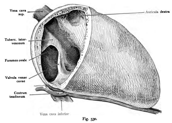
Fig. 530. Fetal heart (6 months) in normal situation
The right atrium is opened. After the alcohol-Semp Terpentinmethode seen dried.
The vena cava inferior vena cava passes through the foramen in the dorsal
Scope of the right atrium. The valvula venae cavae (Eustachii) leads the
Blood according to the foramen ovale into the left atrium. The superior vena cava enters
through the ventral wall of the atrium, and their blood flows and ventral
medially by the foramen ovale into the right ventricle. Below the valvula venae
cavae, the mouth of the coronary sinus with the valvula sinus coronary (Thebesi).
- This text is a Google translate computer generated translation and may contain many errors.
Images from - Atlas of the Development of Man (Volume 2)
(Handatlas der entwicklungsgeschichte des menschen)
- Kollmann Atlas 2: Gastrointestinal | Respiratory | Urogenital | Cardiovascular | Neural | Integumentary | Smell | Vision | Hearing | Kollmann Atlas 1 | Kollmann Atlas 2 | Julius Kollmann
- Links: Julius Kollman | Atlas Vol.1 | Atlas Vol.2 | Embryology History
| Historic Disclaimer - information about historic embryology pages |
|---|
 |
Reference
Kollmann JKE. Atlas of the Development of Man (Handatlas der entwicklungsgeschichte des menschen). (1907) Vol.1 and Vol. 2. Jena, Gustav Fischer. (1898).
Cite this page: Hill, M.A. (2024, April 27) Embryology Kollmann530.jpg. Retrieved from https://embryology.med.unsw.edu.au/embryology/index.php/File:Kollmann530.jpg
- © Dr Mark Hill 2024, UNSW Embryology ISBN: 978 0 7334 2609 4 - UNSW CRICOS Provider Code No. 00098G
Fig. 530. Fetales Herz (6. Monat) in natiirliclier Lage.
Der rechte Vorhof ist geöffnet. Nach der Semp ersehen Alkohol-Terpentinmethode getrocknet.
Die Vena cava inferior tritt durch das Foramen venae cava in den dorsalen Umfang des rechten Vorhofes. Die Valvula venae cavae (Eustachii) führt das Blut nach dem Foramen ovale in den linken Vorhof. Die Vena cava superior tritt ventral durch die Wand des Vorhofes ein und ihr Blut strömt ventral und medial vom Foramen ovale in die rechte Kammer. Unterhalb der Valvula venae cavae ist die Mündung des Sinus coronarius mit der Valvula sinus coronarii (Thebesi).
File history
Click on a date/time to view the file as it appeared at that time.
| Date/Time | Thumbnail | Dimensions | User | Comment | |
|---|---|---|---|---|---|
| current | 23:34, 16 October 2011 | | 733 × 524 (83 KB) | S8600021 (talk | contribs) | {{Kollmann1907}} |
You cannot overwrite this file.
File usage
The following 2 pages use this file: