File:Kingsbury1922 plate03.jpg: Difference between revisions
m (→Plate 3) |
mNo edit summary |
||
| Line 27: | Line 27: | ||
{{Footer}} | {{Footer}} | ||
[[Category:Neural]][[Category:Historic Embryology]][[Category:1920's]] | [[Category:Neural]][[Category:Historic Embryology]][[Category:1920's]] | ||
[[Category:Chicken]] | |||
Latest revision as of 14:08, 22 November 2019
Plate 3
Median plane reconstructions from sagittal sections of the chick embryo, head region only. The notochord is indicated by cross—barring, the neural plate stippled, entoderm and preaxial mesoderm (prechordal plate) stippled; the ectoderm shown in black.
anterior portion, X 67. To illustrate the ventral end of the ‘sutura terminalis marking the anterior end Of the brain—plate.
All figures at the same magnification, X 50.
27.Series 139, 6—7 somites.
28. Series 106, 10 somites.
29. Series 119, 14 somites.
30. Series 109, 16 somites.
31. Series 127, 22 somites.
32. Series Gage 54s, 30 + somites.
33. Ventral View of a model of the head of a chick, eight to nine somites, Back of it is the ‘hypophyseal area and a shallow Seessel’s pocket continuous caudally with a dorsal pharyngeal groove.
Reference
Kingsbury BF. The fundamental plan of the vertebrate brain. (1922) J. Comp. Neural. 461-490.
Cite this page: Hill, M.A. (2024, May 8) Embryology Kingsbury1922 plate03.jpg. Retrieved from https://embryology.med.unsw.edu.au/embryology/index.php/File:Kingsbury1922_plate03.jpg
- © Dr Mark Hill 2024, UNSW Embryology ISBN: 978 0 7334 2609 4 - UNSW CRICOS Provider Code No. 00098G
File history
Click on a date/time to view the file as it appeared at that time.
| Date/Time | Thumbnail | Dimensions | User | Comment | |
|---|---|---|---|---|---|
| current | 13:52, 22 November 2019 | 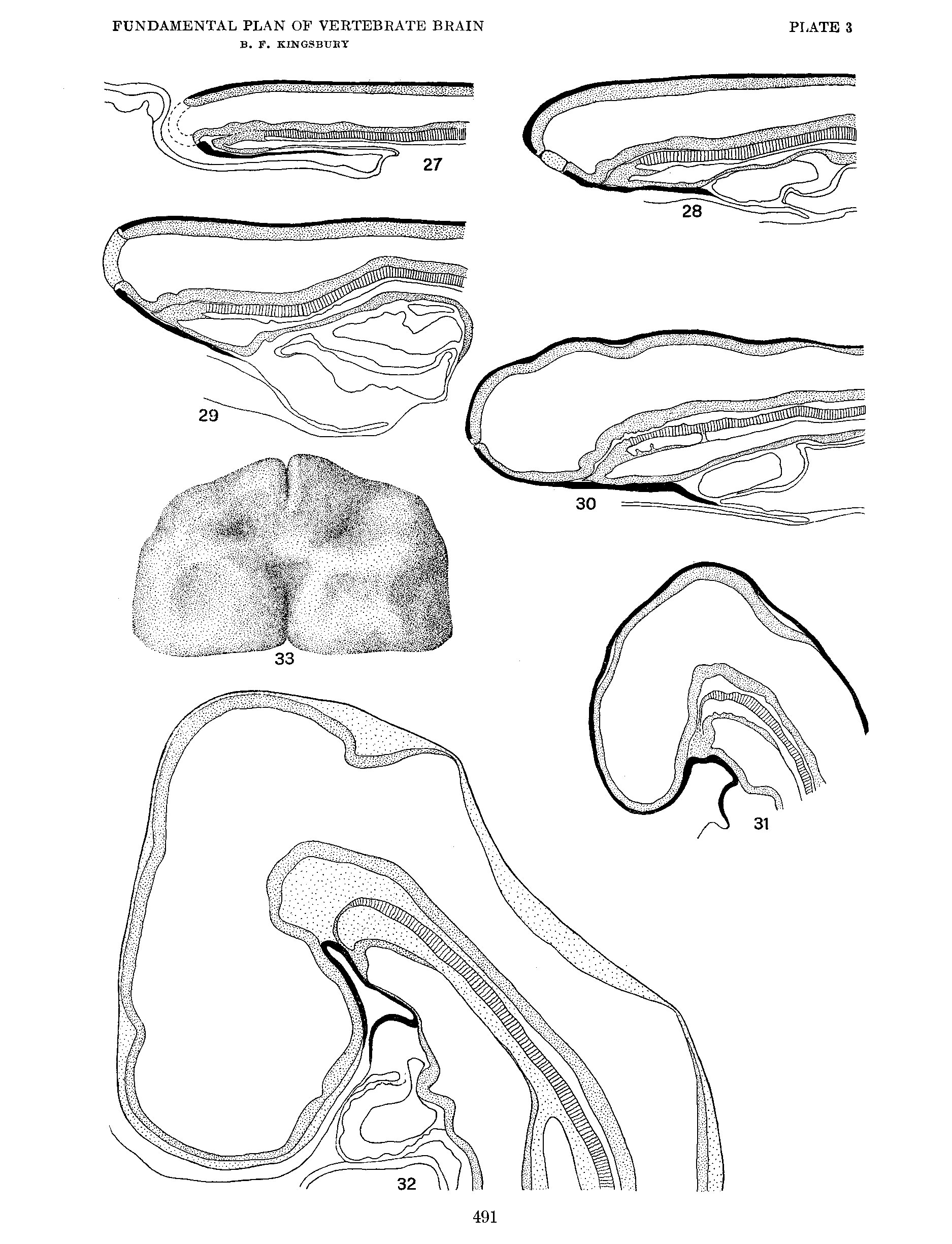 | 1,875 × 2,450 (743 KB) | Z8600021 (talk | contribs) | {{Ref-Kingsbury1922}} |
You cannot overwrite this file.
File usage
The following page uses this file: