File:Kingsbury1922 plate02.jpg
Original file (1,875 × 2,273 pixels, file size: 573 KB, MIME type: image/jpeg)
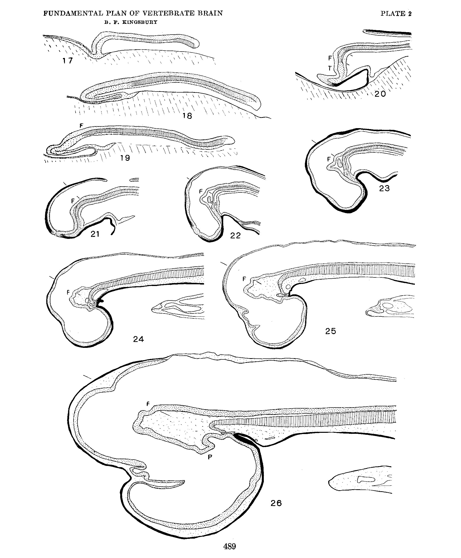
Plate 2
Median plane reconstructions from sagittal sections of Squalus acanthias. The notochord is indicated by cross—barring, the neural plate stippled, entoderm and preaxial mesoderm, stippled or black, ectoderm generally in black. All figures at the same magnification, X 25.
Online Editor - Squalus acanthias = Spiny dogfish; entoderm is the historic term for endoderm.
17. Series no. 42, no somites, length 1.5 mm.
18. Series no. 50, 8 somites, length 2.7 mm.
19. Series no. 52, 11-12 somites, length 2.9 mm.
20. Series no. 55, 15 somites, length 4.0 min.
21. Series no. 56, 16 sornites, length ca. 3.0 mm.
22. Series no. 51, length 4.5 mm.
23. Series no. 70, 28-30 sornites, length ca. 5.0 mm.
24. Series Gage 3, length 9-10 mm.
25. Series no. 46, length 9-10 mm.
26. Series no. 38, length 23 mm.
F, actual or prospective location of the fovea isthmi and the anterior end of the floor plate.
P, Tuberculum posterius (v. Kupffer).
I, Tubercle of the floor (see text).
Reference
Kingsbury BF. The fundamental plan of the vertebrate brain. (1922) J. Comp. Neural. 461-490.
Cite this page: Hill, M.A. (2024, April 27) Embryology Kingsbury1922 plate02.jpg. Retrieved from https://embryology.med.unsw.edu.au/embryology/index.php/File:Kingsbury1922_plate02.jpg
- © Dr Mark Hill 2024, UNSW Embryology ISBN: 978 0 7334 2609 4 - UNSW CRICOS Provider Code No. 00098G
File history
Click on a date/time to view the file as it appeared at that time.
| Date/Time | Thumbnail | Dimensions | User | Comment | |
|---|---|---|---|---|---|
| current | 13:51, 22 November 2019 | | 1,875 × 2,273 (573 KB) | Z8600021 (talk | contribs) | {{Ref-Kingsbury1922}} |
You cannot overwrite this file.
File usage
The following page uses this file: