File:Keith1902 fig180.jpg

Original file (872 × 800 pixels, file size: 141 KB, MIME type: image/jpeg)
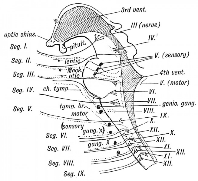
Fig. 180. A Diagram to show the Relationship of the Cranial Nerves to the Primitive Segments of the Head
The Segments to which the Cranial Nerves belong.
1st Cranial Segment — The motor nerve is the 3rd or oculo-motor. The ciliary ganglion, a derivative of the Gasserian, represents the sympathetic ganglion. Ganglion cells representing a vestigial posterior root may be found on the trunk of the nerve. The ophthalmic division of the 5th appears to represent its posterior or sensory root.
2nd Cranial Segment — The motor nerve is the 4th. The sensory is represented by the superior maxillary division of the fifth. Meckel's ganglion represents the sympathetic ganglion. It is known to be derived from the same group of nerve cells as the Gasserian ganglion.
3rd Cranial Segment — The motor nerve is the 6th and motor fibres of the fifth. The sensory root is represented by the inferior maxillary division of the 5th. The otic and submaxillary represent its sympathetic ganglia.
4th Cranial Segment — The motor nerve is the 7th. The sensory root is represented by the chorda tympani and great superficial petrosal, which are developed from the geniculate ganglion (Dixon). The eighth nerve and its ganglia also belong to the sensory system of this segment. The great superficial petrosal represents a splanchnic nerve, the chorda tympani the nerve on the anterior margin of the 1st visceral cleft (see p. 34).
5th Cranial Segment — The motor fibres of this segment have probably been scattered. Some may still remain in the 9th cranial nerve (glosso-pharyngeal) which is the chief nerve of the segment. The ganglia on the trunk of the glosso-pharyngeal represent the posterior root ganglion. The tympanic branch and small superficial petrosal represent an afferent (sensory) splanchnic branch.
6th, 7th, 8th and 9th Cranial Segments — It has been already mentioned (pages 152 and 161) that the four posterior cranial segments are probably trunk segments which have become modified and added to the head. The anterior or motor nerve roots of these four segments are combined in the 12th nerve. Motor visceral fibres, which issue by the anterior roots of spinal nerves, here issue by the vagus and bulbar part of the spinal accessory (all of which are properly designated vagal fibres — Sherrington) and represent the visceral motor fibres of the four posterior cranial segments. The ganglia on the root and trunk of the vagus represent part of a posterior root ganglion. From these ganglia are developed the sensory visceral fibres connected with the fore gut and all the structures derived from the fore gut or splanchnopleure of the fore gut. A vestigial posterior root ganglion may occur on the 12th nerve.
The circuitous course of the spinal accessory is probably due to the migration of the trapezius caudalwards from a cephalic to its present position.
- Brain and Spinal Cord: Fig. 158. Human Neural Tube about 14 days | Fig. 159. Neural Tube Four Primary Divisions | Fig. 160. Zones of the Spinal Neural Tube Gth week | Fig. 161. Zones of the Embryonic Spinal Cord. | Fig. 162. Hind Brain 5th week | Fig. 163. Lateral Neural Tube 5th week | Fig. 164. Inferior Medullary Velum 5th month | Fig. 165. Frog Cerebellum and 4th Ventricle | Fig. 166. Human Cerebellum 3rd month | Fig. 167. walls of the fore-brain | Fig. 168. Human Cerebral Vesicles and Thalamencephalon 3rd month | Fig. 169. Adult 3rd Ventricle | Fig. 170. Fore and Mid-brain 5th week | Fig. 171. Fetal Brain 4th month | Fig. 172. Lamina Terminalis and Primitive Callosal Gyrus | Fig. 173. Corpus Striatum in Cerebral Vesicle | Fig. 174. Cerebral Hemisphere 2nd month | Fig. 175. Cerebral Hemisphere 5th month | Cerebral Hemisphere 7th month | Fiq. 177. Opercula and Fissure of Sylvius | Fig. 178. Ape Island of Reil and Fissures | Fig. 179A. Anthropoid Island of Reil | Fig. 180. Cranial Nerves and Primitive Segments | Figures
| Historic Disclaimer - information about historic embryology pages |
|---|
| Pages where the terms "Historic" (textbooks, papers, people, recommendations) appear on this site, and sections within pages where this disclaimer appears, indicate that the content and scientific understanding are specific to the time of publication. This means that while some scientific descriptions are still accurate, the terminology and interpretation of the developmental mechanisms reflect the understanding at the time of original publication and those of the preceding periods, these terms, interpretations and recommendations may not reflect our current scientific understanding. (More? Embryology History | Historic Embryology Papers) |
Human Embryology and Morphology (1902): Development or the Face | The Nasal Cavities and Olfactory Structures | Development of the Pharynx and Neck | Development of the Organ of Hearing | Development and Morphology of the Teeth | The Skin and its Appendages | The Development of the Ovum of the Foetus from the Ovum of the Mother | The Manner in which a Connection is Established between the Foetus and Uterus | The Uro-genital System | Formation of the Pubo-femoral Region, Pelvic Floor and Fascia | The Spinal Column and Back | The Segmentation of the Body | The Cranium | Development of the Structures concerned in the Sense of Sight | The Brain and Spinal Cord | Development of the Circulatory System | The Respiratory System | The Organs of Digestion | The Body Wall, Ribs, and Sternum | The Limbs | Figures | Embryology History
Reference
Keith A. Human Embryology and Morphology. (1902) London: Edward Arnold.
Cite this page: Hill, M.A. (2024, April 27) Embryology Keith1902 fig180.jpg. Retrieved from https://embryology.med.unsw.edu.au/embryology/index.php/File:Keith1902_fig180.jpg
- © Dr Mark Hill 2024, UNSW Embryology ISBN: 978 0 7334 2609 4 - UNSW CRICOS Provider Code No. 00098G
File history
Click on a date/time to view the file as it appeared at that time.
| Date/Time | Thumbnail | Dimensions | User | Comment | |
|---|---|---|---|---|---|
| current | 11:34, 18 January 2014 |  | 872 × 800 (141 KB) | Z8600021 (talk | contribs) |
You cannot overwrite this file.
File usage
The following 3 pages use this file:
