File:Keibel1897 plate01.jpg: Difference between revisions
m (→Fig. 1) |
m (→Fig. 2) |
||
| Line 9: | Line 9: | ||
Full exact proportion can not be determined at this embryo because the rearmost end of the primitive streak is handled. | Full exact proportion can not be determined at this embryo because the rearmost end of the primitive streak is handled. | ||
===Fig. 3=== | |||
The embryo, which is illustrated in FIG. 3 and 3a, was the mother animal taken 15 days one hour after mating and fixes with Pikrinschwefelchromsäure. I could see in the review of 3 on each side and each Urwirbelpaare accrued before and after yet another without limit cranial resp. caudal. Fig. 3 shows the ring intersected the chorion to the embryo, and you can see how the chorionic turns into the amnion. Only in the districts in which the amnion is still open, you can see directly on the dorsal side of the embryo, otherwise you see the P ^ mbryonalkörper only by the chorion shine through. The caudal end of the embryo is hook-shaped bent downwards. This embryo is the Kopfamnion less developed than the Schwanzamnion, cranial run the two lateral amniotic folds acute angle against each other; caudal they go in the bow into one another; by the still fairly wide opening of the Amnionnabels you see on the medullary and the somites. | |||
{{Keibel1897 figures}} | {{Keibel1897 figures}} | ||
Revision as of 16:05, 22 June 2015
Plate 1.
Fig. 1
The embryonic disc p.7 b. 93 is from a 14-Days 10 hours before the slaughter sow occupied. After the embryonic disc was removed in the usual way with chrome acetic uterus, it was fixes to sublimate on. FIG. 1 illustrates the embryonic disc is how they presented itself to me after this treatment in 70 "Alcohol. M., and M, are a draft after ZiKGi.ER'schen model, which was produced with a basis Plattenreconstruction. We see, as the primitive streak has already been limited to the rear half of the embryonic disc. Before the primitive streak region we see the first installation of the medullary and the medullary. The medullary groove extends up close to the front end of the embryonic disc, rear end already on the point, the front end to embrace the primitive streak. The area in front of the front end of Primitiv.streifens is easy aufgewulstet, and this bead forms the base of the rear part, the medullary groove. At the front end of the embryonic disc a little bump already at surface observation stands right in front of the medullary groove clearly emerges. As can be seen from the average series, called these bumps, the front end of the head extension resp. the notochord, it corresponds to the front end of the embryo at all. The small protrusion that is noticeable at the rear end of the embryonic disc is caused by Mesodermwucherung and the mesodermal Allantoisanlage. It should be emphasized that in this embryonic disc, the posterior intestinal bay is already well trained. The occurrence of the amnion was already mentioned in the previous embryonic disc, the development of the amnion has made progress, we find, as in the previous stage, evenly developed in circumference around the embryonic disc.
Fig. 2
If the germinal disc shown in FIG. 2, the medullary groove in the fore significantly compared to the primitive streak. Even more clearly than in the previous stage it can be seen as the front gewulstete end of the primitive streak is encompassed by the Medullarfalten. Forward towards the medullary forks before the hump, which corresponds to the front end of the notochord and at all the embryo developed. The head of the embryo starts to stand out.
Full exact proportion can not be determined at this embryo because the rearmost end of the primitive streak is handled.
Fig. 3
The embryo, which is illustrated in FIG. 3 and 3a, was the mother animal taken 15 days one hour after mating and fixes with Pikrinschwefelchromsäure. I could see in the review of 3 on each side and each Urwirbelpaare accrued before and after yet another without limit cranial resp. caudal. Fig. 3 shows the ring intersected the chorion to the embryo, and you can see how the chorionic turns into the amnion. Only in the districts in which the amnion is still open, you can see directly on the dorsal side of the embryo, otherwise you see the P ^ mbryonalkörper only by the chorion shine through. The caudal end of the embryo is hook-shaped bent downwards. This embryo is the Kopfamnion less developed than the Schwanzamnion, cranial run the two lateral amniotic folds acute angle against each other; caudal they go in the bow into one another; by the still fairly wide opening of the Amnionnabels you see on the medullary and the somites.
| Historic Disclaimer - information about historic embryology pages |
|---|
 |
Reference
Franz Keibel, Normentafeln zur Entwicklungsgeschichte der Wirbelthiere (Normal plates of the development of vertebrates) Volume Hft.1 (1897) Jena, G. Fischer, Germany.
Cite this page: Hill, M.A. (2024, May 3) Embryology Keibel1897 plate01.jpg. Retrieved from https://embryology.med.unsw.edu.au/embryology/index.php/File:Keibel1897_plate01.jpg
- © Dr Mark Hill 2024, UNSW Embryology ISBN: 978 0 7334 2609 4 - UNSW CRICOS Provider Code No. 00098G
File history
Click on a date/time to view the file as it appeared at that time.
| Date/Time | Thumbnail | Dimensions | User | Comment | |
|---|---|---|---|---|---|
| current | 09:49, 20 November 2013 | 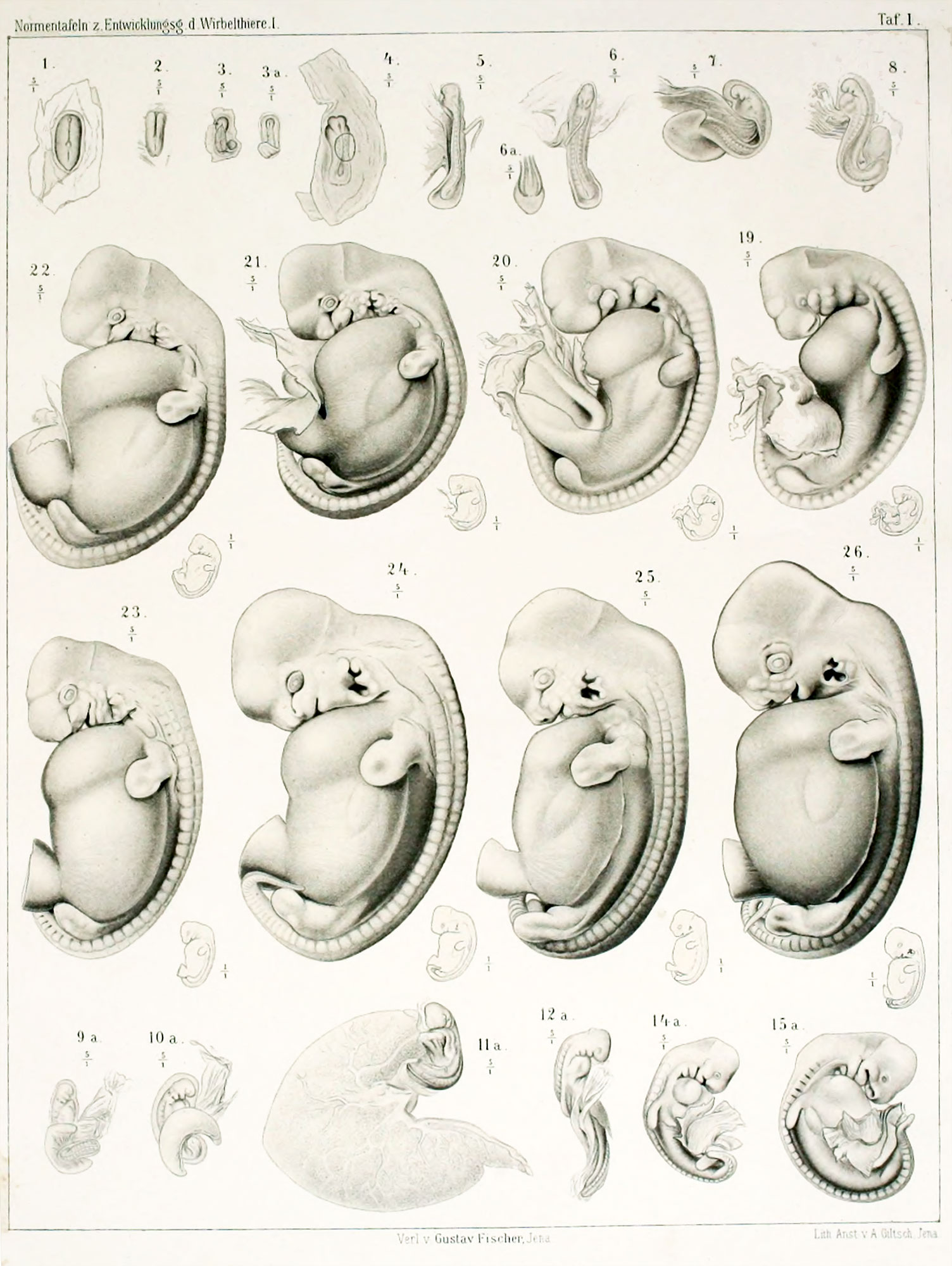 | 1,504 × 2,000 (365 KB) | Z8600021 (talk | contribs) |
You cannot overwrite this file.
File usage
The following 3 pages use this file: