File:Jordan1918 fig04.jpg

Original file (586 × 871 pixels, file size: 78 KB, MIME type: image/jpeg)
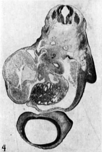
Fig. 4. Section just below the point of bifurcation of the trachea
Section at approximately level a—a’ in figures 2 and 3 just below the point of bifurcation of the trachea. This is the point where the single-cell ‘angioblasts’ (hemoblasts) and the smaller cell-clusters (‘blood-islands’) begin to appear ventrally in both aortic roots. The section shows also the base of the right fore limb-bud, the right brachial plexus, the esophagus, the left ventricle, the bubus cordis, the inferior vena cava, the left duct of Cuvier, the liver with the ductus venosus dorsally, and the telencephalon with the right olfactory placode. The celom contains considerable blood at the right. Photo X 15. (In comparing the photograph of this section with fig. 2 the top should be turned to the right; with fig. 3, to the left.) (In the process of paraffin embedding the specimen changed its shape somewhat, chiefly through aceentuation of the several flexures, so that it no longer corresponds exactly with the form shown in the photographs 1 to 3, in consequence of which the level of sections cannot be indicated with absolute precision in the latter, nor any longer quite accurately with straight lines. Previous to embedding the embryo had been stained in tote with Delafield’s hematoxylin. It was sectioned at 10 microns.)
| Historic Disclaimer - information about historic embryology pages |
|---|
| Pages where the terms "Historic" (textbooks, papers, people, recommendations) appear on this site, and sections within pages where this disclaimer appears, indicate that the content and scientific understanding are specific to the time of publication. This means that while some scientific descriptions are still accurate, the terminology and interpretation of the developmental mechanisms reflect the understanding at the time of original publication and those of the preceding periods, these terms, interpretations and recommendations may not reflect our current scientific understanding. (More? Embryology History | Historic Embryology Papers) |
- Links: Fig. 1 | Fig. 2 | Fig. 3 | Fig. 4 | Fig. 5 | Fig. 6 | Fig. 7 | Fig. 1-6 | Jordan 1918 | Historic Embryology Papers
Reference
Jordan HE. A study of a 7 mm human embryo; with special reference to its peculiar spirally twisted form, and its large aortic cell-clusters Volume 14, Issue 7, pages 479–492, July 1918
Cite this page: Hill, M.A. (2024, April 27) Embryology Jordan1918 fig04.jpg. Retrieved from https://embryology.med.unsw.edu.au/embryology/index.php/File:Jordan1918_fig04.jpg
- © Dr Mark Hill 2024, UNSW Embryology ISBN: 978 0 7334 2609 4 - UNSW CRICOS Provider Code No. 00098G
File history
Click on a date/time to view the file as it appeared at that time.
| Date/Time | Thumbnail | Dimensions | User | Comment | |
|---|---|---|---|---|---|
| current | 10:02, 8 November 2015 |  | 586 × 871 (78 KB) | Z8600021 (talk | contribs) |
You cannot overwrite this file.
File usage
The following page uses this file:
