File:Jordan1918 fig01.jpg
Jordan1918_fig01.jpg (593 × 561 pixels, file size: 44 KB, MIME type: image/jpeg)
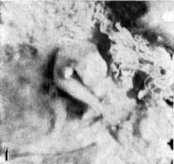
Fig. 1. View of embryo from right side, attached to chorion
It is sharply curved about the neck region, so that the tip of the forcbrain touches the umbilicus. It is also twisted through a complete spiral, the mid-dorsal line coming into view caudal to the right fore limb-bud, and the left hind limb-bud showing below the pelvic flexure. The rhombencephalon, the eye, and the right vena capitis lateralis are also conspicuous; and certain of the nuchal and pelvic myotomes may be seen. Approximately thirty-eight somites could be counted. The chorionic villi are seen beyond the reflected edges of the opened vesicle. This is the View obtained when the embryo was turned on the short umbilical cord so as to expose the side opposite to the one shown when the embryo was in its more natural position as in figure 2. In this position it could only be maintained by force. When detached from the chorionic vesicle, by cutting from the wall of the vesicle a square piece of tissue including the area of attachment of the umbilical cord, the embryo assumed the position shown in figure 3, when the right cephalic surface was turned uppermost.
Photo. X3. All photographs were made by Mr. F. L. Foster.
| Historic Disclaimer - information about historic embryology pages |
|---|
 |
- Links: Fig. 1 | Fig. 2 | Fig. 3 | Fig. 4 | Fig. 5 | Fig. 6 | Fig. 7 | Fig. 1-6 | Jordan 1918 | Historic Embryology Papers
Reference
Jordan HE. A study of a 7 mm human embryo; with special reference to its peculiar spirally twisted form, and its large aortic cell-clusters Volume 14, Issue 7, pages 479–492, July 1918
Cite this page: Hill, M.A. (2024, April 28) Embryology Jordan1918 fig01.jpg. Retrieved from https://embryology.med.unsw.edu.au/embryology/index.php/File:Jordan1918_fig01.jpg
- © Dr Mark Hill 2024, UNSW Embryology ISBN: 978 0 7334 2609 4 - UNSW CRICOS Provider Code No. 00098G
File history
Click on a date/time to view the file as it appeared at that time.
| Date/Time | Thumbnail | Dimensions | User | Comment | |
|---|---|---|---|---|---|
| current | 10:01, 8 November 2015 | | 593 × 561 (44 KB) | Z8600021 (talk | contribs) |
You cannot overwrite this file.
File usage
The following page uses this file: