File:Jordan1918 fig01-06.jpg

Original file (1,383 × 2,320 pixels, file size: 372 KB, MIME type: image/jpeg)
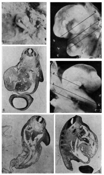
Fig. 1-6. Human Embryo 7 mm
Fig. 1. View of embryo from right side, attached to chorion. It is sharply curved about the neck region, so that the tip of the forcbrain touches the umbilicus. It is also twisted through a complete spiral, the mid-dorsal line coming into view caudal to the right fore limb-bud, and the left hind limb-bud showing below the pelvic flexure. The rhombencephalon, the eye, and the right vena capitis lateralis are also conspicuous; and certain of the nuchal and pelvic myotomes may be seen. Approximately thirty-eight somites could be counted. The chorionic villi are seen beyond the reflected edges of the opened vesicle. This is the View obtained when the embryo was turned on the short umbilical cord so as to expose the side opposite to the one shown when the embryo was in its more natural position as in figure 2. In this position it could only be maintained by force. When detached from the chorionic vesicle, by cutting from the wall of the vesicle a square piece of tissue including the area of attachment of the umbilical cord, the embryo assumed the position shown in figure 3, when the right cephalic surface was turned uppermost. Photo. X3. All photographs were made by Mr. F. L. Foster.
Fig. 2. Left side-view of embryo. The left ventricle and the bulbs cordis are conspicuous. The anlages of the cerebral hemispheres, the eye, the rhombencephalon, three branchial arches, the umbilical cord, the hind limb-buds and the
tail are also clearly outlined. The photograph was retouched so as to bring into
sharper relief also the fourth branchial arch, the left fore limb-bud and the otocyst
with the short ductus endolymphaticus. Magnification X 8. The lines a—a’,
b—b’, and c—c’ indicate approximately the levels of sections in figures 4 to 6.
Fig. 3. View of right cephalic surface. The mass to the left consists of amnion above, and fused amnion and chorion below. The right fore limb-bud has become pushed forward by the spiral twisting and turned so as to bring its originally ventral border on a line parallel with the ventral surface of the pharynx. Just above the branchial arches may be seen through the translucent tissue the vena capitis lateralis. Photo X 8.
Fig. 4. Section at approximately level a—a’ in figures 2 and 3, just below the point of bifurcation of the trachea. This is the point where the single-cell ‘angioblasts’ (hemoblasts) and the smaller cell-clusters (‘blood-islands’) begin to appear ventrally in both aortic roots. The section shows also the base of the
right fore limb-bud, the right brachial plexus, the esophagus, the left ventricle,
the bubus cordis, the inferior vena cava, the left duct of Cuvier, the liver with
the ductus venosus dorsally, and the telencephalon with the right olfactory placode. The celom contains considerable blood at the right. Photo X 15. (In
comparing the photograph of this section with fig. 2 the top should be turned to
the right; with fig. 3, to the left.) (In the process of paraffin embedding the
specimen changed its shape somewhat, chiefly through aceentuation of the several
flexures, so that it no longer corresponds exactly with the form shown in the
photographs 1 to 3, in consequence of which the level of sections cannot be indicated
with absolute precision in the latter, nor any longer quite accurately with straight
lines. Previous to embedding the embryo had been stained in tote with Delafield’s hematoxylin. It was sectioned at 10 microns.)
Fig. 5. Section approximately at level b~b’, figs. 2 and 3, showing left forelimb-bud below. (To compare with fig‘. 2 the top of the figure should be turned to left; with fig. 3, to right.) Five spinal ganglia are shown; also the mesonephros, with the pest-cardinal vein dorsally. The section passes through the point where the yolk-stalk was attached to the primitive ileum (shown in transverse section as minute opening at extreme tip of mesentery). In the lower portion of the mesentcry are shown the vitelline arteries (superior mesenteric artery). This portion of the cclom contains considerable blood. The aorta is completely filled with blood. Within the umbilical cord, to left of mesentery, is shown the umbilical vein. Adjacent sections contain the largest of the aortic cell-clusters (fig. 7). Photo X 15.
Fig. 6. Section approximately at level c—c’, figs. 2 and 3, through the length of the umbilical cord, showing point of reflection of amnion onto cord, below at the left. (To compare with fig. 2, turn top of figure to left; with fig. 3, to the right). The celom contains much blood; the blood-cells are perfectly preserved and entirely normal. On either side of the umbilical cclom are shown the large umbilical veins. The ventral ramus of the aorta is the inferior mesenterie artery. Note the compressed notochord; it is as wide as in fig 5, Where the section passes very obliquely. Photo. X 15.
| Historic Disclaimer - information about historic embryology pages |
|---|
| Pages where the terms "Historic" (textbooks, papers, people, recommendations) appear on this site, and sections within pages where this disclaimer appears, indicate that the content and scientific understanding are specific to the time of publication. This means that while some scientific descriptions are still accurate, the terminology and interpretation of the developmental mechanisms reflect the understanding at the time of original publication and those of the preceding periods, these terms, interpretations and recommendations may not reflect our current scientific understanding. (More? Embryology History | Historic Embryology Papers) |
- Links: Fig. 1 | Fig. 2 | Fig. 3 | Fig. 4 | Fig. 5 | Fig. 6 | Fig. 7 | Fig. 1-6 | Jordan 1918 | Historic Embryology Papers
Reference
Jordan HE. A study of a 7 mm human embryo; with special reference to its peculiar spirally twisted form, and its large aortic cell-clusters Volume 14, Issue 7, pages 479–492, July 1918
Cite this page: Hill, M.A. (2024, April 28) Embryology Jordan1918 fig01-06.jpg. Retrieved from https://embryology.med.unsw.edu.au/embryology/index.php/File:Jordan1918_fig01-06.jpg
- © Dr Mark Hill 2024, UNSW Embryology ISBN: 978 0 7334 2609 4 - UNSW CRICOS Provider Code No. 00098G
File history
Click on a date/time to view the file as it appeared at that time.
| Date/Time | Thumbnail | Dimensions | User | Comment | |
|---|---|---|---|---|---|
| current | 09:28, 8 November 2015 |  | 1,383 × 2,320 (372 KB) | Z8600021 (talk | contribs) | '''Fig. 1.''' View of embryo from right side, attached to chorion. It is sharply curved about the neck region, so that the tip of the forcbrain touches the umbilicus. It is also twisted through a complete spiral, the mid-dorsal line coming into view ca... |
You cannot overwrite this file.
File usage
The following page uses this file:
