File:Johnston1907 fig020.jpg

Original file (1,280 × 1,632 pixels, file size: 277 KB, MIME type: image/jpeg)
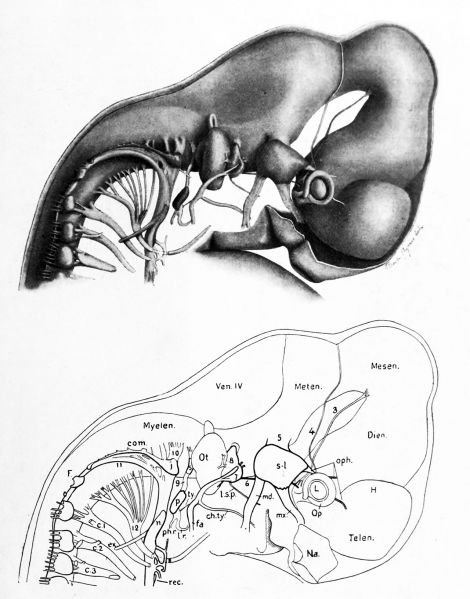
Fig. 20. Reconstruction of the brain and cerebral nerves in a 12 mm pig embryo
(From Minot's "Laboratory Text-Book of Embryology" Drawn and revised by F. T. Lewis.)
Nerves. 3, oculomotor. 4, trochlear. 5, trigeminal, with its semilunar ganglion, s-l, and three branches, oph., ophthalmic; mx., maxillary; md., mandibular. The motor portion, which goes with the mandibular branch, is concealed in this view by the sensory portion. 6, abducens. 7, geniculate ganglion of the intermedius. Fibers from this ganglion mix with the motor fibers of the facial nerve, so that both sensory and motor fibers are found in the three branches, /. s. p., large superficial petrosal; ch. ty., chorda tympani; /a., facial. 8, acoustic, showing an upper vestibular portion, and a lower cochlear portion. 9, glossopharyngeal with its superior ganglion, s, above; its petrosal ganglion, p, below; and its three branches, ty., tympanic; /. r., lingual ramus; ph. r., pharyngeal ramus. 10, vagus, with its jugular ganglion, /, extending posteriorly as a ganglionic commissure, com.; and below, its ganglion nodosum, n. Its branches form the laryngeal plexus,beyond which is the recurrent nerve, rec. Just below the jugular ganglion is the auricular branch of the vagus. II, accessory, which joins the vagus; ex., its ramus externus. 12, hypoglossal. P., Froriep's hypoglossal ganglion. C. I, C. 2, C. 3, cervical nerves.
Brain and Sense Organs. Telen., telencephalon. Dien., diencephalon. Mesen. r mesencephalon. Meten., metencephalon. Myelen., myelencephalon. H., hemisphere. Ven. IV., roof of the fourth ventricle. Op., optic cup. L, lens. Na., nasal pit. Ot., otocyst.
Johnston JB. The Nervous System of Vertebrates. (1907) Blakiston's Son & Co., London.
| Historic Disclaimer - information about historic embryology pages |
|---|
 |
Cite this page: Hill, M.A. (2024, April 28) Embryology Johnston1907 fig020.jpg. Retrieved from https://embryology.med.unsw.edu.au/embryology/index.php/File:Johnston1907_fig020.jpg
- © Dr Mark Hill 2024, UNSW Embryology ISBN: 978 0 7334 2609 4 - UNSW CRICOS Provider Code No. 00098G
File history
Click on a date/time to view the file as it appeared at that time.
| Date/Time | Thumbnail | Dimensions | User | Comment | |
|---|---|---|---|---|---|
| current | 22:58, 23 February 2020 | | 1,280 × 1,632 (277 KB) | Z8600021 (talk | contribs) | |
| 22:53, 23 February 2020 | 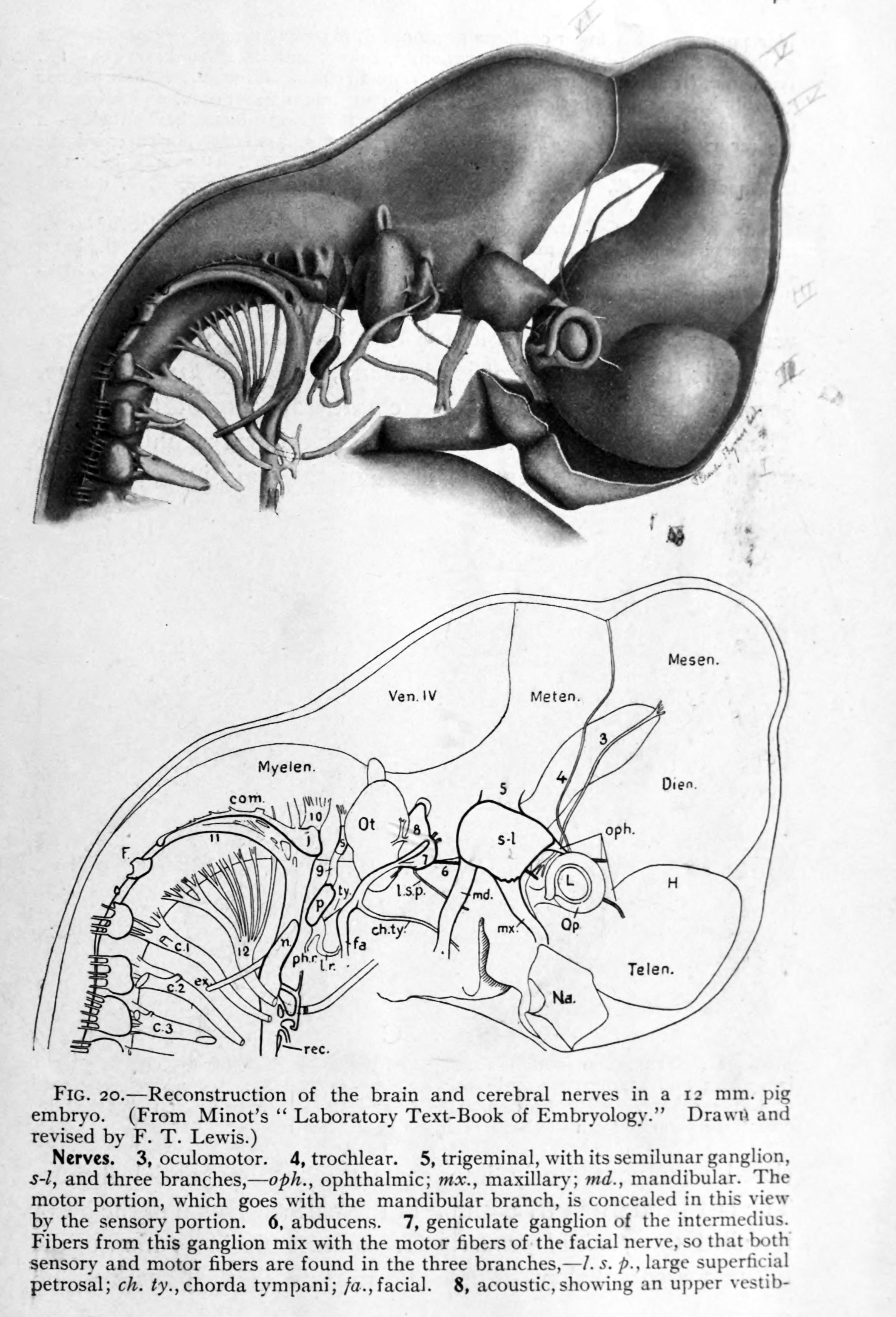 | 2,328 × 3,421 (849 KB) | Z8600021 (talk | contribs) | FIG. 20. Reconstruction of the brain and cerebral nerves in a 12 mm. pig embryo. (From Minot's " Laboratory Text-Book of Embryology." Drawrt and revised by F. T. Lewis.) Nerves." 3, oculomotor. 4, trochlear. 5, trigeminal, with its semilunar ganglion, s-l, and three branches, oph., ophthalmic; mx., maxillary; md., mandibular. The motor portion, which goes with the mandibular branch, is concealed in this view by the sensory portion. 6, abducens. 7, geniculate ganglion of the intermedius. Fibe... |
You cannot overwrite this file.
File usage
The following 2 pages use this file: